Site sections
Editor's Choice:
- Diseases of the musculoskeletal system and connective tissue
- Topic: White Blood Cell Physiology
- Daily teach to help and be grateful for help yourself
- Khachapuri with cheese - the best recipes
- The postulates of successful breeding sheflera at home
- Growing cyclamen from seeds
- What honey is not sugared: varieties, causes
- Symptoms, course, treatment and prevention
- Lecture Topic: Digestive System
- How many grams of honey in a teaspoon and a tablespoon
Advertising
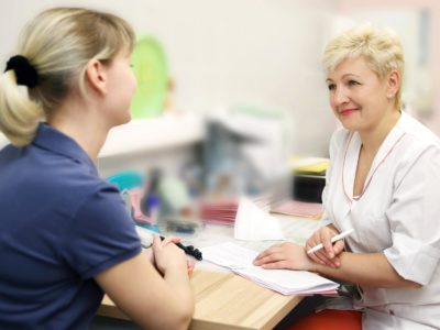
| Diseases of the musculoskeletal system and connective tissue. Orthopedics |
|
The musculoskeletal system in the human body is responsible for the ability to make movements, without which life would be quite limited. Together with tendons, muscles and cartilage, the bone skeleton supports all internal organs, provides upright posture and protects against external damage. The musculoskeletal system, along with other parts of the body, is exposed to all kinds of diseases. Most of them can be successfully treated even at the initial stage. The main thing is to see a doctor on time. Anatomical dataIn total, the musculoskeletal system includes 206 bones, the union of which is called the skeleton. Next, muscles and soft tissues join it, which contain nerve fibers and blood vessels. It is thanks to this structure that the correct course of metabolic processes is achieved, whose role in the life process is difficult to overestimate. As a result, special cells are also formed - osteoblasts, which reliably protect bones from damage and disease, participating in their growth and healing during fractures. Certain areas of the musculoskeletal system and connective tissue are more often than others exposed to excessive, sometimes uncontrolled physical exertion, infectious and independently acquired inflammatory processes, teenage distortions and even displacements. If the problem is not started, then it is easily eliminated under the supervision of an experienced doctor. Neglect also causes further deformation of the musculoskeletal system and connective tissue, the occurrence of severe pain with subsequent impaired motor function. The problem can be felt at absolutely any age. Therefore, even if you feel slight discomfort, the body must be examined. What can you face?Diseases of the musculoskeletal system are quite diverse, so making a diagnosis on your own is a rather ungrateful and very dangerous business. This should be done only by a professional who thoroughly knows the specifics of the structure of the musculoskeletal system and connective tissue.
In this case, the main treatment is directed precisely at this problem. If the problems with the musculoskeletal system were the result of another disease, then first you have to deal with it, and only then deal with the accompanying problems. As a rule, congenital diseases are the most difficult to treat, but this does not mean at all that they need to be left to chance. The minimum that can be done is to alleviate the patient's condition by acting on the muscles and bones with drugs or physiological procedures. A positive result may not be immediately noticeable, but it will definitely be! Not a single person on the planet is safe from injuries, so you have to learn to deal with their consequences. Sometimes it can be very difficult, but diseases must be treated. Even if it takes several years. The consequences of injuries usually make themselves felt for a long time. Oncological processes rarely occur in the area of \u200b\u200bthe musculoskeletal system and connective tissue. They usually get there as metastases. Fighting cancer, of course, is very difficult, but under the supervision of an experienced specialist, this can be quite real. The main thing is faith and perseverance. Musculoskeletal system - one of the strongest systems of human organs. The musculoskeletal system is a skeleton for the human body and allows direct walking. The skull consists of 8 pairwise connected bones. They must be perfectly symmetrical. The bones of the skull can move and move. The skull protects the human brain from physical damage and strictly repeats its shape. Displacement of the bones of the skull at birth can cause cerebral palsy, strabismus and hearing loss. This may be due to displacement of the mother’s pelvic bones. After birth, gradually become harder, but still, they can be corrected. The spine consists of 7 cervical, 12 thoracic and 5 lumbar. If you look at a person in profile, you can see that there are 2 deflections. 1 in the neck and 1 in the lumbar region. Deflection of the spine allows you to remove the load from the vertebral discs. Between the vertebral discs there is a pulpous nucleus, which acts as a shock absorber. Inside the cervical vertebrae are openings through which the cervical veins and cervical arteries pass. This connection provides cerebral circulation. The habit of sleeping on a high pillow and any movements that displace the cervical vertebrae for a long time or injure them can impair blood circulation and cause chronic diseases and stroke. At the bottom of the spine, hip joints are attached to the sacrum. The natural deflection of the spine in the lower back allows you to remove the load from it, shifting the center of gravity from it. If you remove the deflection - the sacrum will wedge into the pelvic bones. Excessive stress will lie on the spinal discs, which will ultimately lead to injury. The pelvic bones should be at the same level. The habit of wearing weights on one side of the body, using only one leg as a supporting leg, can cause mixing of the pelvis. The displacement of the pelvic bones can be determined by the displacement of the level of the shoulders. With the left shoulder raised - “shorter”, that is, the right leg will be higher. And vice versa. This causes a general asymmetry of the body, and as a result, heart pains, mastopathy, and kidney problems can occur. All that is numb or sore in the muscular system, if it is not associated with direct injuries, is most often the result of general asymmetry of the body and pinched current, pinched nerves coming from and to the spine. Scoliosis is not a condition of the spine, it is a condition of the pelvic bones that act as the foundation. When a person constantly shifts body weight on one side, the foot bends greatly. In order for a person not to fall on the thumb, a bone grows, then there will be cartilage and compaction. If a bone has grown on both legs, this means that the person first stood on one leg, then it began to hurt, and he began to stand on the other. You can even your pelvis with gymnastics and manual therapy. Inflammatory processes in the joints, if there were no injuries, this is a consequence of diseases. Under each joint are lymph nodes. If the body is affected by any infection (streptococcus, chlamydia, etc.), thrombosis of the lymph nodes occurs. Fluid will accumulate and collect in the joint. If the body lives for a long time or lymphatic filtration occurs, that is, every day along with the infection, the cartilage that the infection infected will thin out. In this case, you need to go through an antibacterial, antiviral and antifungal program. The fluid in the joint should be clean. The quality of the joint fluid can be determined by the state of the nails. Nails are frozen joint fluid that flows out and hardens evenly every day. Nails should be clear and firm. If the nails are grooved, then something is wrong with the liquid. If the fungus on the nails - the same fungus is in the joint fluid, and it is worth treating the whole body from a fungal infection. If the nails exfoliate - there is a deep violation of mineral metabolism, plus the remains of the fungi are washed out. White dots on the nails are not digested proteins. Impaired protein metabolism means that proteins are not absorbed or digested. If there are white or slightly pinkish stripes on the nails, this can indicate poisoning with salts of heavy metals and pollution of the liver. If there is cervical osteochondrosis, the joint fluid will leak unevenly, and accordingly, tubercles on the nails will form. The spine must be treated starting with the bones of the pelvis. Chinese or Japanese gymnastics would be a good choice. Strabismus and crooked teeth may be the result of uneven bones of the skull. This text will be replaced The musculoskeletal system of Butakov video 1. Ecology.Does not affect. 2. Food.A lack of calcium, silicon, phosphorus, sulfur, or amino acids can affect. The diet should be complete. 3. Water.In its absence, the vertebral discs dry out, elasticity decreases. 4. Psychology.Does not affect 5. Injuries.They are of great importance. Damage to one of the elements of the system causes others to adjust. Harmony is broken. 6. Heredity.Deformed bones can be inherited, especially teeth, since the shape of the skull and the proportions of the face are transmitted. Scoliosis is not inherited. The appearance, the internal structure, and not the disease, is transmitted. 7.Medicine.Does not affect. They are the cause of inflammatory processes. One of the most powerful is psoriatic arthritis. 9. The movement.It has a very strong influence. It is necessary to observe the correct technique for performing exercises and avoid unsuccessful postures. It is advisable to sleep only on the back and with a roller under the neck. Musculoskeletal Recovery Algorithm
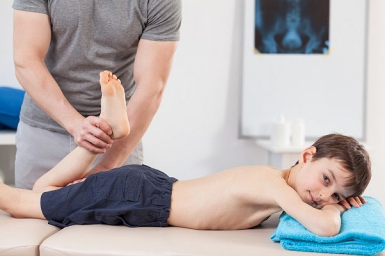
As a result of the trauma, bone deformation occurs. Infringement of blood vessels and nerve conductors occurs. After a concussion, nausea can be felt throughout life. Injury to the back of the skull can result in loss. There are visual buffs that can be pressed by an unadjusted bone. After injury, strabismus may appear. The eyes located in the orbits of the orbits will adjust. The brain will do the same. In case of injury to the cervical vertebrae, the arteries responsible for the blood supply to the brain will be pinched. The same thing can be obtained if you do not sleep properly. When the vertebral artery is pinched below the ears, a noise or ringing in the ears will be heard. Blood pressure will be heard. In order for the neck to straighten, it is necessary to correct the base of the musculoskeletal system - the pelvic joints. And then train your muscle corset. - A disease that is an inflammatory process in the joint tissue as a result of infection. It is generally accepted that arthritis is a disease exclusively of the elderly. But, unfortunately, it is often found in children. Causes of arthritis in children:
The disease develops gradually, therefore, certain difficulties arise in its diagnosis. Symptoms of Arthritis in ChildrenIf the baby still does not know how to talk, you need to more closely monitor his behavior and well-being. With the development of arthritis, he has such ailments:
The disease is a lesion of the joints and cartilage. It is quite difficult for children to carry, because the bones at their age have not yet become stronger, so some of their parts are cartilage. CausesThe most common causes of arthrosis in a child are:
These causes contribute to the deformation of joints and cartilage, which leads to arthrosis. SymptomsFor children's arthrosis, the following symptoms are characteristic:
There are several stages of the disease. The early stage is easily treatable. In the later stages, irreversible deformation of the cartilage is observed. Therefore, treatment should be started as soon as possible.
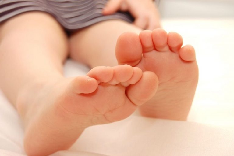
Flat feet- A fairly common childhood illness. It is not a serious illness, but if improperly treated, it can lead to unpleasant consequences. Reasons for the appearanceThere is no single reason for the development of the disease. There are several factors that contribute to the occurrence of flat feet, namely:
For the correct formation of the arch of the foot, the child should be given the opportunity to walk without shoes in the warm season. SymptomsAttentiveness to your baby will quickly suspect that he has flat feet. The most striking manifestations are:
If the child has these symptoms, you need to seek the advice and diagnosis of an orthopedist.
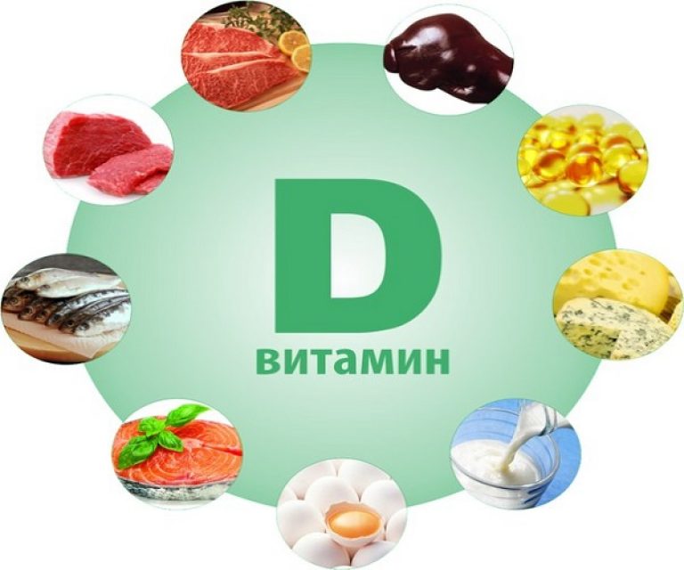
Rickets- A disease in which damage to the bones and central nervous system occurs. Associated with a lack of vitamin D in the body. Rickets are commonly observed in rapidly growing children. The reasons for the development of rickets in a child:
The main reason is the lack of vitamin D in the baby's body, which is the main component for the normal development of bone tissue. Symptoms of the diseaseRickets is most common in children under 1 year old. The baby has such signs:
During the period of active development of rickets, such additional ailments appear:
This disease delays the normal physical development of the baby. He begins to sit or walk much later than his peers.
ScoliosisScoliosis is a fairly common disease in modern children. Usually scoliosis affects school children. The disease is a pathological curvature of the spine. CausesThis disease can be observed in many modern children. Most often, the prerequisites for its development are:
SymptomsWith the development of scoliosis, the child begins to experience such ailments:
In order to avoid diseases of the musculoskeletal system in a child, it is required to provide the baby with the necessary amount of vitamins and regularly visit a pediatrician for preventive examinations. Clinical manifestations of diseases of the musculoskeletal systemPAIN Pain is the most common symptom of this group of diseases. Stimulation of peripheral receptors by irritating agents causes pathological patterns of nerve impulses, which is interpreted in the higher centers of the brain as pain. The manifestation of pain is altered by analgesics, as well as neurohormones called endorphinswhich work as endogenous pain blocking substances. For pain, localization, prevalence, irradiation or orientation are described. Localized pain occurs on the side of the pathological process, while diffuse pain appears when the deeper tissues are affected. Radicular pain occurs due to the anatomical location of the peripheral nerve, due to damage to the nerve or its root as sciatica. The pain radiates away from the pathological focus. Examples of radiating pain include knee pain as a manifestation of a disease of the joint of the big toe or secondary pain in the buttocks with pathology of the spine. Muscle pain occurs due to direct damage to the muscles or their spasm with the formation of reflex muscle contracture. Peripheral nervous pain can be with external (neuralgia) or internal compression of the nerve between anatomical formations (compression neuropathy), with ischemia, infection (herpes zoster), metabolic or toxic (e.g. arsenic) disorders. Bone injuries of the spine, such as cancer metastases, multiple myeloma, infection, or a compressive fracture due to osteoporosis also produce persistent local and rarely radicular pain. Pain in the upper limbs.Shoulder pain occurs when the intervertebral disc protrudes in the cervical spine forward in the presence of a tumor, cuff rupture of the rotator muscle or impingement syndrome (reflected pain), with arthritis of the shoulder joint. Pain can also be directed to the shoulder from the heart, lungs, or pleura. A rotator muscle has a common tendon, represented by supraspinatus and infraspinatum, subscapularis and small round muscle. Tendonitis and bursitis are the result of the process spreading from the rotator muscle to the coracoid ligament (impingement syndrome). Cuff rupture of the rotator muscle is accompanied by acute pain and difficulty in active abduction of the shoulder. The diagnosis is made by X-ray examination of the joint, MRI or ultrasound. Pain in the elbow is associated with epicondylitis (“tennis player's elbow”), compression of the ulnar nerve in the joint, inflammation of the elbow joint, radicular pains arising from compression of the nerve root. Pain in the wrist or wrist is associated with "inflammation of the tendon - the" trigger finger ", or de Quervain's disease. Pathology of the cervical discs.This disease can cause radicular or neck pain; neurological symptoms and symptoms of compression of the spinal cord or roots of the spinal nerves. Weakness in movement or sensory impairment arises from the distribution of the radicular nerves or diffusely below the level of damage that can be seen on the myelogram or MRI. Most often C 4 -C 6 is affected, acute pain occurs, the presence of neurological symptoms depends on the level of damage and the length of the hernia. Compression of the spinal cord will cause weakness during movement and increased reflexes in the lower extremities, in rare cases, urinary incontinence. Treatmentconsists in fixing and traction of the cervical spine, the use of anti-inflammatory drugs. If traction does not regress neurological symptoms, then surgical discectomy at the appropriate level is necessary. Pain in the lower limbs.The most common causes of foot pain are associated with the plantar surface and the metatarsus. It occurs with repeated loads on the heads of the metatarsal bones or at the site of attachment of the plantar ligament to the calcaneus. Additional causes of pain in the foot and ankle joint are caused by joint inflammation, subluxation of the fibular tendon, stress fracture and tarsal tunnel syndrome. Repeated loads on the tibia can lead to a condition such as “shin in a longet” or to pain that restrains motor activity, to soreness over the tibia and stress fractures. Muscle cramps, deep vein thrombosis, ischemia or lumbar radicular syndrome, radiating due to disc herniation or stenosis of the spinal canal, can also cause leg pain. Pain in the knee joint is caused by tearing of the meniscus, inflammation of the joint, chondromalacia of the patella and inflammation of the tendon of the patella or pathology of the thigh. Pain in the thigh appears with adversity in the groin and is noted with arthritis, osteonecrosis, synovitis, tumors or infections. Pain in the lateral thighs is associated with trochanteric bursitis. For pain in the posterior thigh, sciatica and pathology of the lumbar spine should be differentiated. Lower back pain Lumbar syndromeoccurs with a disease or damage to the lumbosacral spine and can be acute or chronic. In the table. 41.1 lists the causes of lower back pain. Lumbar clinically, pain can lead to impaired motor activity as a result of arbitrary restraint of movements, may be aggravated by sneezing or coughing, combined with muscle spasm. The pain extends to the buttocks or legs, compression of the roots causes radicular pain and / or symptoms of impaired movement and sensitivity along the nerve roots. Table 41.1. Causes of Low Back Pain 1. Structural (abnormal or transitional vertebrae, spina bifida, or spina bifida, spondylolysis, spondylolisthesis, anomalies of the articular surfaces)
Examination of the patient includes determining the severity of impaired movement, a test for raising the straightened leg (to irritate the sciatic nerve; if the test is positive, then radicular pain) and a complete neurological examination. The development of lumbar syndrome is associated with traumatic or mechanical causes, and the expectation of the effect of conservative therapy (bed rest, anti-inflammatory drugs, locally warm, in rare cases, antispasmodics for muscle spasm, followed by mobilization, isometric back and exercises for the abdominal press, in rare cases corset or bandage for the back) is quite natural. Spondylolisthesis- subluxation of the body of one vertebra over another. Reasons: 1) defect of a part of the joints, 2) fracture of the posterior elements, 3) congenital deficiency of the articular surfaces, 4) insufficiency of the articular surfaces due to degenerative diseases of the intervertebral discs, 5) lengthening of the intervertebral space. Clinically, spondylolisthesis may be asymptomatic, although pain in the back, hamstring, sciatic nerve, and pain of varying intensity and rarely focal neurological symptoms are noted. In the lateral x-ray, the displacement and defect of the vertebra are more visible. Treatmentconservative (rest, physical exercises to strengthen the abdominal press), with the progression of the symptoms of subluxation, which usually occur in the posterolateral connection, is operational. The task of the operation is to eliminate the spondylolisthesis and to restore the anatomical relationship. Sciatica.This is a symptom, not a disease, used to describe radicular pain. It can occur with compression of the roots of the nerves due to a hernia of the intervertebral disc, a tumor, an abscess or narrowing of the hole in degenerative arthritis with hypertrophy of the articular surfaces. In rare cases, the cause is determined by a tumor in the pelvis and buttocks or an abscess, as well as inflammation and toxic damage. Disc herniation or degenerative arthritis are not the most common causes. Hernias appear at the border of L 5 / S, and L 4 / L 5, when there is a rupture of the fibrous ring behind the spinal canal. Violation of the position of the disc causes compression of the roots, radicular pain (sciatica), back pain, sometimes neurological symptoms. In children and the elderly, herniated discs occur infrequently, mainly this is the lot of the middle-aged group. The pain is worse when sitting, coughing, sneezing and bending the legs. Symptoms occur when raising a straight leg, partially with the addition of dorsal flexion of the foot. Damage can be seen on a CT scan, but a clearer image is obtained on a ~ myelogram or on an MRI scan. Treatmentbegin with conservative measures, in 80-90% of patients spontaneous improvement is noted, surgical care is not required. Surgical treatment consists in removing the bulging part of the disk or its enzymatic cleavage by percutaneous injections of chimopapain or collagenase. New approaches to treatment - microdisectomy and percutaneous discectomy - remain controversial. Stenosis of the spinal canal.This is a narrowing of the size of the canal itself or the exit openings of the spinal nerves, which can be acquired with degenerative diseases of the discs or congenital pathology, for example achondroplasia. Diagnosis is by CT or MRI scan. Patients experience pain in the back or leg, aggravated by standing and walking and subsiding in a sitting position, in contrast to pain of discogenic origin. There are neurological complaints and symptoms, including a decrease in reflexes. Treatmentconservative with physical exercises to bend and wear a bandage, "with the introduction of anti-inflammatory drugs or the introduction of steroids into the epidural space. For relapses, wide posterior surgical decompression with or without joint fragments is used. Purulent osteomyelitis of the spine.The most common causative agent of this pathology is staphylococcus aureus,spreading hematogenously from other foci of infection. Patients suffer from back pain, with an X-ray examination, disk destruction is detected, sometimes neurological symptoms occur. Fever or other common signs of infection are possible, but not necessary. Blood leukocytosis, increased ESR. Treatmentconsists of intravenous antibiotics, immobilization and often surgical treatment using a bone graft on the front surface (anterior fusion). MUSCULAR PATHOLOGY Muscle paralysis and muscle spasticity Motor paralysis -it is a loss of voluntary control over muscle contraction. A healthy muscle has some tone during rest, which is absent with damage to the lower motor neurons, which causes flaccid paralysis. There are also no tendon reflexes from the interrupted pathway from the lower motor neuron. Muscle spasticityarises due to pathologically increased muscle tone with passive tension, which is associated with damage to the upper motor neuron. Loss of blocking control of tendon reflexes from the superior motor neuron when it is damaged also causes an increase in reflexes. The electrodiagnosis of disorders of the muscles and nerves is manifested during electromyography (needles are inserted into the muscles to record electrical potentials from them) and the determination of the speed of nerve conduction (electrical stimulation of the peripheral nerve with the recording of the activity of muscle potentials). The clinical degrees of muscle dysfunction are presented in table. 41.2. Table 41.2. Clinical degrees of muscle dysfunction
Congenital muscle disease Muscular dystrophy.A group of hereditary diseases with progressive muscle degeneration. Types: Duchenne (irreversible), fascial-scapular-humeral (benign) and lower limb belts. Myotonia.Hereditary progressive pathology, including myotonic dystrophy and congenital myotonia. Polio.Acute viral disease of the central nervous system, arising from the destruction of cells of the anterior horns of the spinal cord with the formation of flaccid paralysis. In the acute phase, fever, malaise, and headache are noted. At this point, the process may end or, in a few days, the paralytic phase begins to develop, after which the recovery phase begins. Some improvement in motor function may continue for two years. Treatmentconsists in applying physiotherapy to the joints, increasing the volume of passive movements and preventing contractures. In the stage of residual effects, surgical stabilization of the atonic joints by arthrodesis, alignment of limb lengths and tendon transplantation to restore lost functions may be required. Tendons are transplanted at the 1st degree of muscle disorders, at the 4th and 5th degrees only muscles are transplanted. Usually there is a deformation of the foot, corrected first by strengthening, and then by a combination of tendon transplant and arthrodesis with the full development of the skeleton. Cerebral paralysis.There are 3 cases per 100,000 live births. Occurs with a head injury or birth injury, hypoxia or viral diseases (such as measles or cytomegaly). 50% of patients have spastic, 25% - athetoid, 5% - ataxic, 5% - rigid forms and 15% - a mixed clinical picture. 60% of patients with a spastic clinic develop hemiplegia (upper and lower extremities are involved on one side), paraplegia (lower extremities) and tetraplegia (all extremities are affected). Treatmentit is aimed at the prevention of contractures, training in walking and surgical correction of deformities developing in case of impaired muscle function. Strengthening muscles helps prevent equinovarus deformation of the ankle joint (clubfoot), resulting in hip flexion contracture and knee flexion contracture. Adjuvant contracture can cause subluxation and hip dislocation and is painful, which requires treatment by advancing tenotomy, obturator nerveectomy, or varus / derotational osteotomy of the proximal femur. Recently, a transplant of the rectus femoris muscle with a tendon has been used to eliminate the swinging phase of the gait. A correction of the Achilles tendon may be required to correct contracture; monitoring of spinal displacement in progressive neuromuscular scoliosis is rarely needed. Myelodysplasia (spinal dysraphia).This is a defect in the development of the spinal column in combination with neurological disorders. Insufficiency of closure of vertebral elements can be without involvement of the spinal cord (latent spina bifida) or with myelomeningocele, when there is a neural tube defect at the same level. Antenatal screening for yards of a-fetoprotein helps in the diagnosis. Recent scientific evidence suggests that adding folic acid during the first trimester of pregnancy can reduce the risk of myelomeningocele in a baby. In 80% of cases, this pathology is combined with hydrocephalus. Patients with myelomeningocele suffer from paralysis below the site of the defect, and treatmentshould be performed immediately after birth by surgical closure of hernial protrusion and bypass surgery to reduce hydrocephalus. The prognosis for restoration of function depends on the level of damage. With pathology below L 4, the patients are on an outpatient basis, although there are deformities of the lower extremities, such as persistent equinovarus deformity of the foot and subluxation of the thigh, which are corrected by surgery. Violation of sensitivity causes the formation of pressure sores which complicates recovery.
For effective treatment, the following is necessary.
2. Normal joint mobility and muscle strength. 3. Control of the cerebral cortex for voluntary muscle movements. 4. Normal muscle tone, including when changing position. 5. The absence of sensory impairment. 6. Control of the cerebellum for muscle action, balance of visual and auditory mechanisms. Gait disorders occur with neurological distress, for example, with ataxia (damage to the cerebellum, posterior pillars) or with spastic paraplegia (damaged superior motor neuron or cerebral palsy). Mechanical causes include differences in limb length, hip joint pathology (congenital dysplasia or dislocation, hip subluxation, Legg-Calve-Perthes disease, arthritis), knee joint disease (arthritis, dissecans osteochondritis, valgus or varus deformity). A new method for correcting the difference in limb length is called distraction osteogenesis(Ilizarov technique), in which a cortical osteotomy is performed with a gradual bone extension and fixation in the apparatus. Spinal deformities Kyphosis -this is an increase in the normal bulge posterior to the thoracic spine with the involvement of several vertebral bodies. The hump is an acute kyphotic angular deformity that can be congenital after injury or due to infection (tuberculosis). Kyphosis is muscle, in violation of posture (benign and is treated by physical exercises) or discogenic. The latter type has a tendency to progress, combined with abnormalities of the vertebral end plate and herniated disc. Treatment:physical exercises, in rare cases with severe deformities with strengthening of the spine (spinal fusion). The occurrence of senile kyphosis is usually associated with osteoporosis, multiple compression fractures and sphenoid deformity of the vertebrae. Scoliosis.Scoliosiscalled any lateral deviation of the spine from its usual location. Congenital scoliosis is combined with vertebral abnormalities (hemivertebral, full or partial fusion of vertebral bodies). The progression of this type of scoliosis is treated by early limiting the influence of the vertebrae. Paralytic or neuromuscular scoliosis is usually combined with a long S-shaped torus-coryumbal curvature and often requires instrumental intervention if strengthening does not prevent progression. Idiopathic scoliosis progresses during the puberty, and the onset of age and the side of the curvature are important factors. Thoracic curvature under the age of 10 years has a poor prognosis. Progress is minimal after the complete development of the skeleton. The most frequent curvature is the thoracic region to the right side, found mainly in girls. JK lateral curvature is added rotation of the vertebral bodies. The rotational component is emphasized by bending, lateral asymmetry, protrusion of the ribs or transverse processes of the convex side in the direction of curvature. A lateral x-ray shows a flexion curve. Treatmentconsists in a regular x-ray examination to identify the progression of the process and in mandatory physical exercises. With progression, reinforcement or plastic thoracolumbosacral correction is used. An alternative treatment may be electrical stimulation of the muscles on the curvature side, but the results are controversial. When the curvature progresses by more than 40 °, surgical correction with shafts (sub-plate circular fixation) and reinforcement at the back are indicated. Segmental fixation avoids postoperative immobilization. Recently, more rigid fixation has been used to improve the correction of sagittal and lateral curvature. Front reinforcement is used for rigid thoracolumbar curvature or degenerative scoliosis in adults. Deformations of the foot and ankle joint Flat feetmay be plasticor rigid.Congenital plastic flatfoot is the most common type of this pathology, usually painless and benign. Rigid (peroneal spastic) flatfoot occurs as a result of pathology of the tarsus or congenital overlap between the calcaneus and scaphoid, talus or cuboid bones, often painful, can be treated by insolation or resection of the connecting plate. Acquired platypodia occurs due to an injury with a rupture of ligaments in the midfoot, a tendon of the posterior tibial muscle, or impaired muscle function in neurological diseases (polio). CONTRACTS Contracturecalled constant shorteningand rigiditymuscles, joints, and fascial structures that may be congenital or acquired. An example of congenital contractures is clubfoot and multiple deformities. Acquired joint contractures occur after injuries of tissues adjacent to the joint, with impaired muscle function (see previous information on cerebral palsy), with burns or idiopathic causes (Du-Puitren contracture of the palmar fascia). Volkmann's ischemic contracture is a consequence of tunnel muscle syndrome of the forearm after injury. Edema of muscle lacunae is limited to fascia, which leads to ischemia with constant muscle necrosis and lateral fibrosis. Similar problems can occur on the lower extremities. Clinical symptoms are represented by decreased blood circulation or pulse, pain with passive straightening of the affected muscle, paresthesia, and weakness in movement. The diagnosis is made after measuring muscle tunnel pressure using a special catheter and monitor; treatmentconsists in immediate surgical decompression of the affected muscle vagina. Diseases of the pineal gland (osteochondritis or osteochondrosis) Term "Osteochondritisimplies anomalies of the secondary centers of ossification of long tubular bones. Most often, pathological changes are represented by necrosis due to the lack of blood supply to the pineal gland. The most frequent localization of osteochondritis: lunate bone, scaphoid, tarsal-articular joint, epiphyses of the vertebrae, head of the humerus, head of the femur, patella, tuberosity of the tibia, calcaneus and metatarsal heads. Legg-Calve-Perthes disease (Legg-Calve-Perthes) of the thigh often affects boys aged 5-9 years, a bilateral process occurs in 10% of cases. In the initial (or prodromal) stage, patients limp, complain of pain in the thigh or knee. Then, loss of movement in the thigh and flattening of the femoral head (coxa plana) are joined. Later, after the restoration of the circulation of the pineal gland, the symptoms and complaints decrease, although the restriction of movement and deformation of the femoral head may remain constant. Treatmentshould be aimed at holding the head inside the acetabulum and at strengthening in the reduced state. The patient should be transferred to outpatient monitoring until the recovery (or revascularization) stage, which usually takes about 1-2 years, has completely passed. In some severe cases, varus osteotomy is required to hold the head. Osgood-Schlatter disease (Osgood-Schlatter) is a lesion of the tibial tuberosity in patients 13-15 years old, sometimes a history of previous trauma is indicated. Pain, soreness and an increase in the tuberosity of the tibia with the appearance of radiological signs are noted. Treatmentsymptomatic, limitation of activity, in more severe cases - a plastic cylindrical splint or immobilization of the knee joint for 6 weeks. The disease is self-limiting, although the swelling of the tibial tubercle remains constant In Köhler’s disease (Cybeg), the tarsal scaphoid joint in children 3–6 years of age is involved. Pain and swelling are noted. The sclerosis of the scaphoid is visible on the x-ray. Treatment:plastic tire for “several weeks and cast” the arc for subsequent support Congenital orthopedic diseases DEFORMATIONS DETECTED AT BIRTH Adduction of the metatarsus, valgus deformity of the foot, unilateral rotation of the leg, internal tibial torsion and adduction of the thigh with external rotation as a result of improper position in the uterus can cause persistent equinovarus deformation of the foot. Usually, this pathology can be corrected during exercises with passive straightening; tires are rarely required. Congenital dislocation of the femur consists of a partial or complete displacement of the femoral head from the acetabulum, there are 0.67 cases per 1000 live births. Treatment is most successful in the early stages, so all children under one year old should be examined by an orthopedist to exclude this pathology. On examination, the hip clicks - a symptom of Ortolani (Orto-lani), dislocation may occur, abduction and flexion are limited. Symptoms: limitation of abduction by 75 ° or less, undoubted shortening of one hip - a symptom of the Galeazzi (Gale.az.zi), and asymmetry of the gluteal folds. If a small child is not diagnosed, then lameness and an overwhelming gait will be noticeable when walking. An x-ray of the ossification of the pineal gland and the acetabular index (angle between the acetabulum and the horizontal line) are detected more than normal (22 e). With radiological. .MRI and 4 3 studies revealed hip dislocation. Treatmentsubluxation in newborns is carried out by a device for fixing Pavlik (PaVlik) with wearing it for 3-6 months. If the abduction of the thigh does not decrease or the child is large enough in age, then periodic cutaneous traction is necessary, followed by closure with reposition and a spiky bandage on the thigh in the abduction state. An open reposition is necessary for children “older than: one year old or with a late diagnosis. In older children, femoral shortening and osteotomy are important for obtaining persistent concentric reposition with holding the femoral head in the acetabulum. Congenital persistent equinovarus deformity is represented by flexion of the ankle joint, deviation of the foot from normal position, abduction and median rotation of the tibia. There are approximately 4 cases per 1000 live births. Without treatment, the deformation becomes permanent and makes it difficult to move. Treatmentbegin immediately after birth with passive straightening exercises followed by wearing a corrective plastic tire. First fix abduction, then varus "back foot" and only then - equinus. If the “deformity recurs or the correction is incomplete, the surgical release of the“ back foot ”should be with an open reposition of the deformity and subsequent wearing of the tire. If the child is old enough, moderate relapse can be treated with lateral transplantation of the anterior tibial muscle tendon and / or lengthening of the Achilles tendon. Congenital valgus deformity (vertical talus) causes dislocation of the talus-navicular articulation with the vertical location of the talus and the location of the navicular with the back surface of the talus. The sole of the foot has a flat-foot deformation and rigidity. The early use of a plastic tire is useful, but in most cases, surgical reduction and a pin are required to lengthen the Achilles tendon. Children of the older age group are shown triple arthrodesis. Multiple congenital arthrogryposis, or amyoplasia, is combined with the replacement of fibrous tissue at birth, resulting in loss of joint mobility, other deformations (congenital hip dislocation, knee dislocation) occur, the treatment of which is carried out according to the method described above. Sprengel deformity (congenital high scapula) is a consequence of embryonic failure when the scapula is placed in the correct position. In rare cases, the shoulder blade is attached to the spine with a tape of fibrous tissue or cartilage, called brachiovertebral mass.Moderate cases do not need treatment; in more severe cases, surgical intervention is indicated, but with a delay of up to 3-6 years. Klippel-Feil syndrome (Klippel-Feil), or congenital short neck, occurs due to multiple layers in the cervical spine and usually does not require treatment. Congenital torticollis - a one-sided contracture of the sternocleidomastoid muscle, causing the head to tilt to one side, can be post-traumatic with painful swelling from previous muscle deformation. Treatmentconsists in performing straightening exercises. In the absence of the effect of treatment or with late diagnosis, surgical correction of the muscle is indicated. Other congenital deformities include radicular ulnar synostosis (stratification of the proximal radius and ulna), distal ray distortion with subluxation of the radicular ulnar joint and congenital aplasia or dysplasia of the long bones (absence of the radius with radial “oblique” or fibula) . Common bone disease BONE COMPOSITION Organic components: 90% collagen type I; the rest are bone-specific phosphoproteins, proteoglycan, sialoprotein, osteonectin, osteosalcin, and growth factors such as transforming growth p-factor, fibroblast growth factor, and bone morphogenetic proteins. Inorganic components: calcium phosphate in the crystalline form of hydroxyapatite and 8-9% water. Bone cell enzymes: osteoclasts contain collagenase, acid hydrolases and acid phosphatase; osteoblasts contain alkaline phosphatase and collagenase activity. There are two primary forms of ossification (ossification), or deposits of minerals in the tissues of the skeleton. Long tubular bones are formed by mineralization of the cartilage with the subsequent transformation of these tissues into bone. This process is called endochondral ossificationand in addition to embryonic bone formation gives rise to long tubular bones. Secondary centers of ossification of the pineal gland appear during the formation of bone callus after a fracture, which is called intramembranous ossification.To do this, bone mineralization is used directly by osteoblasts without a cartilaginous stage. Bone correction is necessary when osteoclasts cause resorption of the bone, which subsequently forms the entire skeleton by close pair formation of bone by osteoblasts. Due to the content of growth factors in the bone matrix, it can be easily transplanted to stimulate local bone formation (osteoinduction). Osteoblasts retain a new bone on their surface (osteoconduction), which is used to make up for lost. Bone grafts are used for failure to fracture, to stimulate arthrodesis and restore bone segments lost during trauma, infection or tumor. Bone allografts are almost always more effective than non-viable autografts giving antigenic cellular material. Recent studies include the use of vascularized bone grafts, where there are micro-seismic anastomoses that allow for quick engraftment of bone segments; synthetic bone grafts are used, such as hydroxyapatite, demineralized bone matrix, or growth factors (bone morphogenetic proteins). PATHOLOGY OF THE DEVELOPING BONE Achondroplasia.The most common form of dwarfism, combined with the normal size of the body, but with shortened limbs, has an auto-somnomodinant type of inheritance. Hollier's disease (Oilier) (dyschondroplasia).Multiple interruptions of cartilage in the metaphysis lead to the deformation of long tubular bones. Transformation into malignant chondrosarcoma occurs in 15-25% of cases. Multiple exostoses.Autosomal dominantly inherited pathology characterized by multiple growth of cartilage outwards metaphysis on the pelvic and long, tubular bones. Surgical removal is required only in case of symptoms or in rare cases of malignant transformation into chondrosarcoma. Polyostotic fibrotic dysplasia.The disease manifests itself in childhood as a result of dysplastic bone formation by cells resembling fibroblasts in the metaphysis and diaphysis of long tubular bones. Pathological fractures or curvature of the bones that require surgical intervention are possible. Imperfect osteogenesis.Hereditary pathology of type I collagen with several subtypes. Patients have blue sclera, deafness, fragile bones and, therefore, fractures at an early age. Fetal forms are very severe, ending in the death of the fetus. The forms of the disease in young children are less severe; teenage forms denoted as imperfect osteogenesisthe least severe. For the prevention of bone fractures in children, osteotomy and the introduction of an intramedullary rod are used. Osteopetrosis.Congenital osteosclerosis. A rare hereditary disease with a defect in osteoclasts that are unable to regenerate bone. It is characterized by radiological bone compaction, anemia, frequent fractures and infections. In severe cases, a bone marrow transplant is necessary. Meloreostosis.The disease causes regional asymmetric osteosclerosis of the cortical bone layer with the appearance of “drops from a candle”, local pain and fibrosis of the adjacent joint during X-ray examination. EXCHANGE VIOLATIONS Morbus (scurvy).Vitamin C deficiency leads to a defect in the cross-linking of collagen and, as a result, to insufficient strength of the vascular endothelium; subperiosteal hemorrhages occur and the density of calcification of the germ zone increases due to impaired regeneration and bone formation. The introduction of vitamin C gives a quick healing effect. Rickets.Vitamin D deficiency can be the cause of many diseases and occurs when there is a malabsorption of calcium from the intestines. Lack of food and intestinal malabsorption syndrome cause inadequate absorption of vitamin D, while kidney and liver diseases lead to insufficient hydroxylation of vitamin D, necessary for its transition into an active form. Long tubular bones in children become soft and bent, the growth zone expands, the pineal glands are enlarged and painful on palpation. Treatment:vitamin D. Vitamin D-resistant rickets is a hereditary disease that requires significant doses of vitamin D and phosphates to treat bone pathology. Givophosphateae.A rare hereditary disease characterized by low alkaline foefatase activity and urinary excretion of phosphoethanolamine. OsteomalaciaEquivalent rickets in adults associated with impaired vitamin D metabolism (see description above). It is characterized by pathological fractures; treatmentspend vitamin D. Fibrous osteitis(parathyroid osteodystrophy). Multiple lesions of bones and adjacent areas as a result of excessive secretion of parathyroid hormone. With primary hyperparathyroidism, hypercalcemia is noted pathological fractures or curvature of long tubular bones. The parathyroidectomy is the method of choice. Osteoporosis.This condition occurs when there is an inadequate amount of bone, which in another respect is biochemically normal. It is associated with Cushing's syndrome, thyrotoxicosis, long-term steroid therapy, and in postmenopausal women with loss of estrogen. Treatment:calcium preparations and physiological doses of vitamin D (it is necessary to replenish any component during osteomalacia), exercise, if necessary, estrogens, in rare cases, antiresorptive drugs (calcitonin or bisphosphonate). Pituitary disorders.Pituitary pituitary in childhood can cause dwarfism, while hyperfunction leads to gigantism. Hyperfunction in adults (usually caused by pituitary adenoma) causes acromegaly with an increase in the skull, chest, and fingers. Hypothyroidism (cretinism).Delayed ossification leads to a decrease in growth and an asymmetrical, osteochondrosis-like physique, as in the case of Legg-Calve-Perthes disease. The disease can be cured if therapy with thyroid hormones is started up to 1 year of age. Mucopolysaccharidosis,There are 12 types of hereditary metabolic disorders of mucopolysaccharides (glycosaminoglycans), which vary in severity and in various combinations with spinal deformities, mental development disorders, bone pathology, corneal opacification and joint stiffness. Paget's disease(deforming osteosis). Disease of bone regeneration associated with a slow viral infection of osteoclasts. In the early stages, there is excessive osterclastic resorption and vascularization, then the formation of pathological bone and sclerosis with trabecular and cortical thickening occurs. At later stages, dense sclerosis of the bone with fibrous degeneration of the bone marrow is observed. The disease begins at the age of 35-50, pain is noted by about 30% of patients. Pathological fractures and a curvature of the length of the tubular bones occur. The activity of alkaline phosphatase in serum and the level of hydroxyproline in the urine are increased, which correlates with the activity of the disease. To block bone resorption (resorption), diphosphonate or calcitonin is used. Violations of the reticuloendothelial system Fat granulomatosis.It occurs with any violations of fat metabolism, with the accumulation of cerebroside lipoproteins, phospholipids, cholesterol, cerebroside protein. An X-ray examination reveals bone lysis. Eosinophilic granulomatosis.It can occur in childhood as a dense formation on the skeleton or multiple formations. This pathology is combined with hepatosplenomegaly, exophthalmos and diabetes insipidus. Solitary eosinophilic granulomatosis can cause changes in the affected vertebra or pathological fractures of long tubular bones; usually amenable to conservative therapy with a self-limiting process. The most severe form of the disease, characterized in newborns, ends fatally. Systemic forms of eosinophilic granulomatosis are treated with vinblastine and prednisone. Hodgkin's disease.A form of malignant lymphoma, manifested by lysis of the bone and secondary involvement of the bone marrow. Symptomatic lesions are susceptible to radiation therapy, systemic manifestations - chemical therapy. Leukemia.It causes bone damage, most often with lymphoblastic leukemia, in which rarefaction of the zone adjacent to the metaphysis growth zone is detected. Multiple myelomaProliferative malignancy of plasma cells, sharply limiting the "perforation" damage to the bone. The disease affects the skull, ribs and long tubular bones; the diagnosis is made according to a bone marrow biopsy or when a pathological monoclonal immunoglobulin is detected by serum immunoelectrophoresis. Hemolytic anemia.It causes changes in the bone marrow in the spine and skull, “hairs at the end” or “sun rays” appear, especially in the skull. Fractures (see tab. 41.3, 41.4 and 41.5). Fractures -these are deformations or violations of the integrity of the bone arising from an excess of force applied along the length of the bone. Fractures are classified by structure (transverse, spiral, oblique, segmental, comminuted), by localization (diaphyseal, metaphyseal, epiphyseal) and combined with damage to the surrounding skin and soft tissues (open or closed). The pathological fracture line passes through the pathological focus, which changes the strength of the bone in case of metabolic disorders or due to a tumor. Clinical manifestations:pain, swelling, deformation, instability and crepitus, ecchymoses. Diagnosis requires an X-ray examination in at least two projections with mandatory seizure of joints above and below the lesion. Inspectionincludes the search for other injuries, assessment of blood circulation and neurological abnormalities in the affected limb. Open fractures should be urgently examined by an orthopedic surgeon; in the operating room, immediate surgical treatment of the wound should be performed to prevent osteomyelitis. Initial surgical treatment should be performed in the first 6-8 hours after the injury, the wound most often does not close tightly. Dead tissue is removed, the fracture is immobilized with an external fixative or plastic splint. Combined vascular damage requires an early restoration of integrity and treatment, since irreversible muscle ischemia occurs in the first 6-8 hours after an injury. With ischemia, the release of fascial vaginas is necessary for the prevention of tunnel syndrome during limb reperfusion. Fat embolism syndrome is a form of adult respiratory distress syndrome, occurs in some patients, more often with multiple fractures of long bones. It occurs 2-3 days after the injury and is characterized by hypoxemia, confusion, fever and transient petechiae. Treatmentlies in the respiratory support and use of corticosteroids. Nerve damage can be combined with fractures. Clinical manifestations range from neuropraxia (transient, recurring deterioration of nerve function) and axonotmes (axons are crossed, but the nerve sheath is intact and regeneration is possible) to the most severe irreversible forms of neurotmes, in which the nerve is completely crossed. The integrity of the crossed nerve should be restored as quickly as possible (with fixation of the fracture or primary surgical treatment of the wound). Joint injuries must be examined radiographically to rule out intra-articular fractures, which often require surgical treatment. Inspection is important to detect instability of the ligamentous apparatus, which can be cured by immobilization or recovery (with partial damage). Arthroscopy is a valuable method of instrumental examination and treatment of joint damage, most often used in pathology of the knee and shoulder joints. Surgical arthroscopy is used for meniscus ruptures, osteochondral fractures, cartilage degeneration, synovitis, shoulder instability due to edge ruptures in the articular fossa of the shoulder joint or intraarticular articular formations, and ruptures of the anterior cruciate ligament. When comparing fragments with an open surgical method, the duration of the disease and the rehabilitation time are reduced. Fracture treatment Stages of fracture treatment: 1. Acute stage:vigorous treatment of somatic disorders. 2. Stage of inflammation:hematoma necrosis of the edges of the fracture, release of kinins, granulation tissue is formed within two weeks. 3. Stage of recovery:cartilage and bone develop from periosteal and mesenchymal cells; cartilage undergoes endochondral calcification, the membranous bone is formed by osteoblasts on the periphery of the corn by the gradual (over several months) replacement of calcified cartilage with bone. 4. Stage of reconstruction:bone marrow becomes layered due to resorption and formation; the bone restores its shape by reconstruction under the influence of mechanical stress. The duration of the process is from several months to several years. Treatment of cartilage damage. Articular cartilage has a very limited ability to repair. Damage leads to the loss of proteoglycans with an increase in mechanical stress, the death of chondrocytes, deformation and progressive degeneration. Surgical penetration into the bone subchondrally allows you to restore the defect by the formation of fibrochart. Fusion delay and integrity violation Delayed fracture fusion leads to a prolongation of the average recovery period and worsens the outcome. Failure to heal leads to inadequate recovery. Causes of impaired adhesion: excess movement of the injured limb, excessive traction, infections and severe soft tissue ruptures. Treatment:bone grafts or stimulation with electromagnetic fields. Pathological fractures Stressful, or pathological, fractures occur when repeated loads on the bone do not cause damage by isolated forces of the same power. Fractures of this series occur after long walks, jogging and other physical exertion. X-ray signs may not be very pronounced, it is necessary to watch the periosteal reaction. Treatment:immobilization. Epiphyolysis The growth of limbs in length is carried out due to the sprout zone adjacent to the pineal gland of long tubular bones. The zone of temporary calcification is the weakest part of the bone, which is why fractures occur here. Salter-Harris classification of damage to the germ zone is widely used. Type I: a transverse fracture through the metaphysis affecting the germinal zone; good prognosis after closed reposition. Type II: partial fracture through the growth zone, leaving through the bone metaphysis; also a good prognosis with a closed reposition. Type III: longitudinal fracture through the articular surface and pineal gland, then transversely through the metaphysis and growth zone; the prognosis is good only when restoring anatomical ratios. Type IV: longitudinal fracture through the pineal gland, sprout zone and metaphysis; An open reduction is usually necessary, a high risk of impaired limb growth in the future. V type: damage to the growth zone by type of crushing; further high risk of impaired limb growth. Fractured children recover faster than adults. Of great importance for recovery is the magnitude of the angular deformation. However, the difference in limb growth occurs after a fracture; angular deformation can increase as in adults. For treatmentclosed fractures use plastic tires. Complications of this method include: edema, which disrupts circulation and causes limb ischemia; bedsores and neuropraxia. Neurocirculatory disorders when using the tire are treated with a support dressing and a soft pad to reduce pressure. Measurement of fascial pressure should be the rule to rule out tunnel syndrome. An increase in fascial pressure serves as an indication for emergency surgical fasciotomy. Tires are applied with the capture of the proximal and distal joints in relation to the damaged bone. Corset is used for fractures of the spine. External fixation is used for open fractures with damage or loss of soft tissue. The spokes inserted above and below the fracture site are connected with external fixators, stabilize the fracture and give quick access to wounds. Another way to stabilize a fracture is traction. In young children, apply skin-sticky tape, in older children and adults use a transverse percutaneous needle for traction. Electrostimulation through knitting needles or by external application of a ring-shaped electrode in a high percentage of cases contributes to the fusion of fragments when this process is delayed due to bone heterogeneity. Joint diseases The joints consist of hyaline articular cartilage coated with a fibrous capsule. The capsule has a layer of synovial cells that secrete synovial fluid, which improves nutrition and lubrication of articular surfaces. The joint cartilage matrix consists of 40% of type II collagen, 40% of proteoglycan and 20% of glycoproteins as an organic component. Minor collagens (types IX and XI) are in the matrix, as are growth factors (transforming B-growth factor and epidermal growth factor). Normally, synovial fluid contains hyaluronate and up to 200 nuclear cells, glucose and electrolytes. Table 41.3. Fractures and injuries of the upper limbs
Continuation of the table. 41.3
Table 41.4. Fractures and injuries of the lower extremities
Continuation of the table. 41.4
Table 41.5. Fractures and injuries of the joints of the pelvis and spine
Inspection of the joint involves the presence of effusion, redness and soreness, as well as measuring the boundaries of mobility, the thickness of the synovial membrane, temperature. An x-ray examination includes an x-ray in two projections, rarely arthrography, if a meniscus or capsule pathology is suspected. The study of synovial fluid, which is normally straw yellow, helps. If free cells can be counted or crystals are available, then the liquid is cloudy. Against the background of inflammation, viscosity decreases with the breakdown of hyaluronates. Clots with insufficient mucus are also a sign of inflammation. Normally, the synovial fluid contains 100 mg / L less glucose than blood serum, and this gradient increases with a large number of white blood cells. Purulent arthritis,bacterial, or septic, is caused by a bacterial infection and is the result of rapid and irreversible destruction of articular cartilage. Staphylococcus aureus and hemolytic streptococcus are the most common pathogens, but many microorganisms can cause the same damage. Patients with fever, effusion, joint pain during movement, leukocytosis and "increased ESR of the blood. The number of leukocytes in the synovial fluid exceeds 50,000, 90% of which are polymorphic nuclear leukocytes. Pathogens can be identified by staining with Gram or taking for sowing fluid aspirated from the joint. Treatment:drainage, mandatory intravenous administration of antibiotics. Some infections are treated with daily aspiration of joint fluid better than permanent drainage. In case of hip joint infection, drainage is performed immediately to prevent necrosis due to circulatory disorders. Arthroscopically often drain the knee joint. Early movements after the acute manifestations of the infection subside are important to prevent joint stiffness, antibiotics continue to be used for 3-6 weeks. Tuberculosis lesionin the USA it is not very common, but it causes severe destruction of bones and joints. The spine is often involved with the destruction of the adjacent vertebrae, the formation of kyphosis and the formation of an abscess. In peripheral joints, a subchondral bone is destroyed early, while changes in articular cartilage are detected at later stages of the disease. The outcome of the process is fibrous joint ankylosis. The clinical course may be without obvious symptoms, and the diagnosis depends on the selection of the pathogen by biopsy or aspiration. Usually, synovial fluid contains less than 200,000 cells / liter of leukocytes: Patients undergo primary surgical treatment of the affected joint, supporting and specific therapy with three drugs at the same time (isoniazid, etham-butol, rifampicin) from 6 months to 1 year. Surgical arthrodesis of a destroyed joint is required. Tuberculous spondylitis (Pott's disease, Pott) is treated with primary surgical treatment, decompression, a rib is used for transplantation in combination with specific therapy with three drugs. Gonococcal arthritismore common in women. At the beginning of the disease, migratory polyarthralgia is noted, then the localization of pain in one or two joints. The knee, elbow and carpal joints are more often affected. Infection can occur in subacute and chronic forms. Treatment:aspiration of the contents, penicillin. Arthritis Lima(Lyme) is called a spirochete Borrelia burgdorferi,mites are carriers. Skin rashes indicate the onset of rheumatic syndrome, resembling rheumatoid arthritis with mild articular syndrome. There are also cardiac and neurological symptoms. Treatment:penicillin, tetracycline. Rheumatoid arthritis -a systemic disease that affects many organs is especially characterized by proliferative destruction of the synovial membrane of many joints. This is expressed in the destruction of articular cartilage, called pannus(superficial diffuse vascular keratitis), periarticular osteopenia and narrowing of the joint space are radiologically determined. 90% of patients have an anti-y-globulin factor called rheumatoid factorwhich is measured by a latex fixation test. On an x-ray, soft tissue swelling, osteoporosis and periarticular erosion are visible, followed by complete destruction of the joint. Treatmentinvolves the appointment of anti-inflammatory drugs (steroid and non-steroid at the same time), gold salts, methotrexate and antimalarial drugs. Surgery:synovectomy of the affected joint and tenosynovectomy for the prevention of tendon rupture; in the later stages of the disease, joint replacement (hip, knee, shoulder, elbow, carpal, metacarpophalangeal and proximal interphalangeal); resection of the head of the metatarsal bones during deformation of the forefoot; various operations on the brush to eliminate deformations. Osteoarthritis, or degenerative arthritis, -a progressive form of joint degeneration of a non-inflammatory nature. Cartilage damage is expressed in the loss of proteoglycans, which leads to fibrillation of the articular surfaces. Marginal osteophytes are an integral part of the processes of destruction and restoration, the surface of the cartilage begins to expose the subchondral bone. X-ray revealed asymmetric narrowing of the joint space, subchondral sclerosis and cysts, osteophytes. Joints are susceptible to diseases such as trauma, Legg-Calve-Perthes disease, septic arthritis, gout, or hemophilia, which can cause changes characteristic of osteoarthritis. The effusion is minimal, the process progresses slowly. Treatmentconsists in limiting mobility, the appointment of anti-inflammatory drugs, walking assistance; osteotomy corrects the deformation of the joints, at a later date, joint arthrodesis or prosthetics. Complications of total arthroplasty: infections, dangling joints and periarticular heterotopic ossification. According to the latest data, incomplete articular fixation is used. implant, prompted by the long-standing problem of “hanging” prostheses when fixing the bone on the inside. The method is becoming popular, especially among young patients. Dentures have a porous metal shell, which allows the bone to grow into this foreign material. Osteonecrosis the femoral head occurs with trauma, sickle cell anemia or impaired fat metabolism (diseases with the deposition of fats, alcoholism, the use of corticosteroids). Joint surfaces decrease during the revascularization phase when bone resorption and mechanical weakness occur, which leads to the rapid development of degenerative arthritis. For early diagnosis, an MRI scan is required; early surgical treatmentbone transplantation or bone marrow decompression prevents the process. Gout- a disease associated with metabolic disorders - crystallization of urates and laying them around the joints, which is manifested by severe attacks of arthritis. The plus-fa is most often affected - “the ulang joint of the big toe in men older than 30 years. An accurate diagnosis is made by the presence of urate crystals in the aspiration fluid from the joint by polarizing light microscopy. Treatment:colchicine or indomethacin in the acute phase, in chronic - allopurinol to reduce the level of urate in the blood serum. Pyrophosphate Arthritis (pseudogout, or chondrocalcinosis) - episodic arthritis resembling gout, with the most common damage to the knee and carpal joints. Diagnosed by detection of pyrophosphate crystals in the synovial fluid; with x-ray examination - meniscus calcification. Treatment:anti-inflammatory therapy. Hemophilia Arthritis
occurs as a result of acute recurrent hemorrhage in the synovial membrane of the joint, usually after a minor injury. The knee, elbow, shoulder and ankle joints are affected. Contractures develop with thickening of the synovial membrane, deposition of hemosiderin, and progression of destruction of articular cartilage. The essence of preventive treatment -minimized hemorrhage; applying a dynamic support dressing to prevent contractures. In the later stages of the disease, prosthetics or arthrodesis under protective transfusion of factor VIII coagulation system.
Diseases of the synovial membrane Pigmentary nodular synovitis- proliferative inflammatory process of the synovial membrane, the cause of which is unknown. There is a thickening of the membrane and tubercular proliferation, which causes progressive destruction of the joint. The aspiration fluid is brown, as it contains hemosiderin. Treatment:early synovectomy, then arthrotomy or arthroscopy. Synovial Chondromatosis- cartilaginous metaplasia of the synovial membrane with the formation of a mobile foreign body inside the joint. The disease slowly progresses, resembles pigmented nodular synovitis, one joint is affected. Treatment:synovectomy. Toxic, or transient, acute hip synovitis develops in childhood, is similar to septic arthritis or Legg-Cal-ve-Perthes disease. Often occurs after a viral disease, manifested by lameness or a child's refusal to walk. It is necessary to exclude septic arthritis, to conduct a study of aspiration fluid from the hip joint. Treatmentsymptomatic: bed rest, skin traction, protection against stress, while there is a pathology. SLIPPING HEAD OF HIP EPIPHYSIS The condition is typical for adolescents 10-15 years old, more often boys suffer. It is manifested by pain in the thigh and lameness. The pineal gland slides medially or along the back of the femoral neck. The impossibility of internal rotation of the thigh and forced walk are noted. Bilateral process occurs in 25% of cases. Diagnosis: by orthogonal radiography of the thigh. Treatment:the introduction of knitting needles into the focus (with subacute or chronic slipping) or closed reduction and the introduction of knitting needles (with acute process). Chondrolysis is a complication of this pathology, more often in blacks and women, secondary degenerative changes develop in old age. JOURNAL NEUROPATHY (SHARKO DISEASE) Joint Charcotoccurs with denervation of the limb. Neuropathy is caused by damage and dryness of the spinal cord, syringomyelia, leprosy, and diabetes. The process goes through the destruction of the joint as a result of repeated trauma with impaired sensitivity, and this can be very fast and severe, with cross instability and swelling, but minimal signs of inflammation. It is difficult to make arthrodesis, therefore conservative methods are used (wearing an orthopedic apparatus). Musculoskeletal system tumors Primary bone tumors are relatively rare, but there are both benign and malignant. Musculoskeletal tumors are characterized by centrifugal growth from a single node, the presence of a pseudocapsule and the non-growth of anatomical boundaries (bones, muscle fascia, joint capsule, neurovascular bundles). Metastases are often hematogenous to the lungs or bones. For tumors of bones and soft tissues, a system for determining the surgical stage of the process is used (Table 41.6). Table 41.6. Stages of tumors of the lean muscle system
|

| Read: |
|---|
New
- What drugs lower cholesterol
- How to accelerate the healing of a leg fracture
- Plaque formation and composition
- How hair straighteners work
- Technological map of the lesson the structure of seeds of dicotyledonous plants
- Review of Old Testament Sexual Vocabulary
- After how much plaster is removed
- Lady's hand kissing allowance
- Summary of the lesson "first aid for bruises and fractures"
- Food Sources of Ascorbic Acid


