Site sections
Editor's Choice:
- Knee fracture treatment and rehabilitation
- Chicken fillet with porcini mushrooms in a pan
- What drugs reduce cholesterol
- How to accelerate the accretion of a leg fracture
- Plaque formation and composition
- How do irons for hair
- Technological map of the lesson structure of seeds of dicotyledonous plants
- Overview of Old Testament Sexual Vocabulary
- After how much plaster is removed
- Handbook for kissing a woman's hand
Advertising
| Do not bend the knee after a fracture of the patella. Knee fracture treatment and rehabilitation |
|
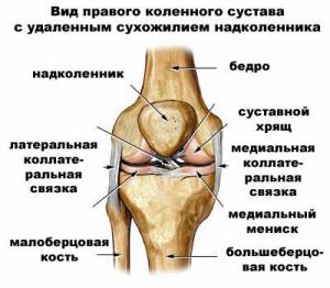
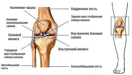
In 50% of cases of injury to the lower limbs, the injury is a fracture of the knee. Legs play an important role in the functioning of the human musculoskeletal system. Exposed to daily loads, legs provide free movement of a person. A serious knee injury is characterized by increased pain, complete immobilization and a long rehabilitation period. The cause of the fracture can be excessive exercise during sports, extreme sports, falling to the knee. In the event of a knee injury due to professional sports activities of a person, the process of treatment and rehabilitation can take a much longer time than with a normal fall. Kinds of injuries and knee fracturesOne of the most difficult and at the same time fragile joints of human bones is the knee joint. In medicine, the definition of "knee joint" means the connection of the fibula and tibial bones with the upper femur. Special meniscus septa connect the above-mentioned parts of the knee to each other. In addition to the menisci, the ligaments, tendons and muscles are involved in the process of joining the bones. Articular cartilage provides a smooth interaction of all components of the knee.
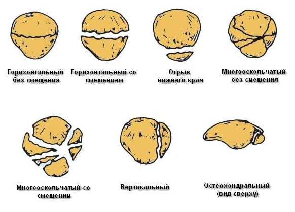
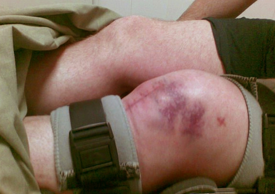
The most common cause of such a fracture is falling on a leg bent at the knee. Fewer cases of fracture occur due to a direct blow to the patella. There are cases in which there was no impact, and the injury was due to an excessively strong strain on the tendons. At the same time the lower part of the patella can come off. The most common horizontal fractures of the knee, often accompanied by displacement of the bones. A blow taken at high speed or with great force may contribute to the formation of a multi-fragment fracture. Individual cases include vertical fractures of the knee. As a rule, they are not complicated by displacement, with rare exceptions. Symptoms of a knee fractureFractured knees correspond to:
First aid for the fracture of the knee jointA key role in the treatment of such injuries is played by first aid to the victim. In the event that the injury is open, the first thing to do is to immediately stop the bleeding. To do this, you must attach ice, a piece of frozen meat or snow to the injured area. Wrap the “cold” in a towel or clothes - low temperatures can damage the skin. In the event that you are alone and there are no people capable of giving you first aid, take the following actions:
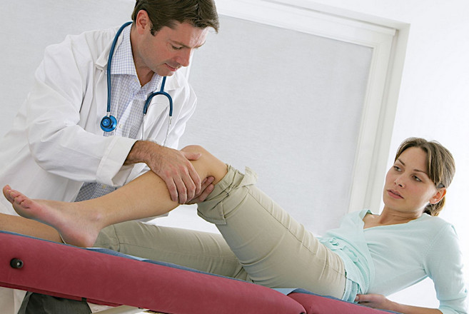
Treatment of fractures and injuries of the knee jointAn ambulance team arrives to send the victim to the hospital. Fractures of the knee joints are classified as the most dangerous injuries and require detailed diagnosis and treatment. Starting with asking the patient, the doctor will proceed to the examination of the injury. Using a method of palpation, the doctor will determine the area of damage, and then prescribe an x-ray. X-ray similar injuries done from several angles, so the traumatologist will examine in detail and examine the fracture site.
After receiving all the comprehensive information about the nature of the fracture, the doctor prescribes treatment. In simple cases of fracture, the injury site will be plastered. Gypsum is applied for at least 1.5 - 2 months. In severe cases of fracture, the doctor prescribes a comprehensive treatment. For complex fractures with significant displacement of the bones and the presence of fragments of bone tissue, surgical intervention may be undertaken. During the operation, the surgeon returns the fractured parts of the knee to their anatomically correct locations and restores the destroyed joint structure. With the help of special devices made of surgical steel, the doctor fastens the particles of the knee. These auxiliary tools include all kinds of pins, metal plates and screws. The process of treatment and recovery of such injuries can be delayed for many months. Rehabilitation periodThe time it takes for the knee to fully recover depends on the severity of the injury. After removing the plaster, the patient may be prescribed the following restorative measures:
It is important to remember that any fracture requires examination and treatment by a specialist.
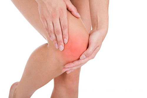
Do not forget that late assistance can lead to disability. Knee fracture is a rather complicated injury, because four bones take part in the formation of this joint. Due to the vulnerability of this area, the knee joint is broken in 10% of cases of the total number of fractures. Moreover, this injury is equally affected by men and women. The age category also varies: from 20 years and more. Symptoms of a knee fractureSigns of a general nature for all injuries of the knee joint should be considered: the identification of acute painful sensations that increase in the process of palpation of the joint, as well as when attempting any, even the most insignificant movement in the area of the knee; systematic formation of puffiness in the knee joint; taking into account the main role of injury in the occurrence and its location, bruising begins to appear; there is a violation of the functioning of the joint, in connection with this, the injured cannot perform any movements of the injured limb, and also cannot step on the affected leg. Causes of knee fractureThe main factors for the occurrence of a fracture of the knee joint are forceful influence on it or significant pressure. The most common fracture of the patella (patella) occurs in the process of falling on the knee, which was in a bent position. More rare are cases of fracture with a direct blow to the patella area, and in some situations a fracture can occur without force. Sometimes, too much traction on the tendon causes injury, which destroys the knee bones and surrounding muscles. With this option, in 90% of cases there is a complete separation of the lower region of the patella, while in the remaining 10% there is a partial separation. Types of knee fracturesFractures in the knee joint are divided into the following categories: thigh injury; trauma to the tibia; injury to the small tibia; patella injury; meniscus injury. In accordance with the division into types, it is accepted to distinguish such fractures of the knee, as the extra-and intra-articular. In the latter case, the rupture of the synovial sac occurs, which covers the outer part of the joint, and in the first variant, only the bone is damaged, without affecting the articular part. Found a mistake in the text? Select it and a few more words, press Ctrl + Enter DiagnosticsAccurate diagnosis of any bones or joints fracture can only be performed using X-ray examination. It is with the help of X-rays that the fracture line is correctly determined, as well as the probable displacement of parts of the knee joint relative to each other. A fracture in the area of the cartilage of the knee joint is characterized by a less vivid clinical picture. It is characteristic of this injury that the painful sensations begin to intensify with the exercise of the movements. In the resting state pain syndrome is not observed. X-ray, in most cases, does not reveal even the most minor deviations. A significant role in the diagnosis of knee injuries is arthroscopy. This procedure involves visualizing the joint area with endoscopic equipment. This method makes it possible to consider a crack in the cartilage tissue, which is the main cause of the impaired optimal functioning of the muscles of the knee joint. Similar to the described clinical manifestations, symptoms are also characteristic of a meniscus fracture, which is determined during arthroscopy. 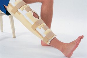 The treatment process should begin immediately after the diagnosis has been made. At the same time, at any stage of therapy, the volume and characteristics of the process may be different, depending on the necessary actions. For example, first aid involves the immobility of the joint by any means available. The purpose of maintaining immobility is the need to prevent further injury to the joint. In addition, at this stage it is desirable to introduce analgesics that prevent pain shock. After immobilization and anesthesia, all actions of the first stage of treatment can be considered complete. The next step in treatment is carried out with the support of a traumatologist. This stage implies the necessity of matching the bone fragments in the anatomically correct order, as well as their fixation in the optimal position. This will help create the most favorable conditions for the healing process. The described mapping can be carried out: closed method, that is, without surgical intervention using manual techniques; open - in this case, the operation is carried out with further fixation of bone tissue with the help of special devices. As a rule, after a fracture of the knee, it is necessary to apply an elastic bandage, which ensures the immobility of the joint. If the articular cartilage has been damaged, at the second stage of treatment it is necessary to use chondroprotectors - medicines that help restore the structure of the cartilage tissue. In some situations it is necessary to use anti-inflammatory agents. For example, a fracture in the area of the condyle of the knee joint, the treatment of which is very often accompanied by the intake of such drugs, in 70% of cases is complicated by autoimmune processes. Anti-inflammatory drugs are used to suppress them. RehabilitationIn the process of treating a fracture of the knee joint, the final stage is rehabilitation, which makes it possible to make the knee motility again full-fledged. Recreational activities include frequent massages, physical therapy, and physiotherapy. All these measures must be assigned directly by a specialist. Only in this case, you can count on a 100% and the fastest possible recovery after such a complex injury as a fracture of the knee joint. 22 Jun 2014 The knee joint is one of the most mobile and multifunctional in the human body, therefore injuries associated with this particular joint are quite common in medical practice. Knee fracture is a complex injury, but with a professional and timely approach, it is completely treatable. There are a great many varieties of injuries of the knee joint, including fractures, since the structure of the knee is a complex mechanism consisting of many components. For a long time, medical scientists have investigated injuries of the knee joint, and at the moment there are several classifications of the types of fractures in this vulnerable area. A special place in them is occupied by intraarticular fractures of the knee joint. Our article about them. Classification of intraarticular knee fracturesFractures of the knee bones inside the joint are divided into two main groups, which in turn are also divided into separate types of damage. Intra-articular fractures of the distal articular femur
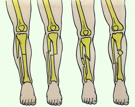
This kind of fractures occur most often in older people, in whom their bone tissue weakens and becomes fragile over time, as well as in young people with large body loads or severe injuries, for example, automobile accidents. Statistics of such fractures suggests possible complications of treatment, such as shortening of the thigh muscles or incomplete restoration of knee function. With such an injury, the bone can break across (transverse fracture) and along (longitudinal fracture), and can also split into several fragments (multiple fracture) or fracture (fractured fracture). Also, fractures of this type can be closed and open. Of particular danger are open fractures of the distal articular end of the femur, as if the integrity of the periarticular muscles, ligaments, tendons and nerve fibers is compromised, complications can occur and the recovery period will be long. Intra-joint fractures of the proximal tibial articular part
This type of fracture can be obtained when falling even from a small height, in particular, from the height of its growth, during car accidents, with undue stress on the knee joint and also with weakening and thinning of the bone tissue caused by diabetes mellitus, malignant tumors or infections. With proper diagnosis, and then with professional treatment, you can return to the joint all its strength, mobility and stability, as well as get rid of all sorts of complications. The methods of treatment are chosen by the doctor depending on the severity of the injury, on the individual characteristics of the patient, on his age and lifestyle. But most importantly, during the rehabilitation period, you should strictly follow the instructions of the doctor, then you can save yourself from the many unpleasant consequences of this fracture. Symptoms and first aid before the arrival of the doctorThe following symptoms may be observed in intraarticular fractures of the knee joint:
Self helpIt is necessary to take some actions before the arrival of the ambulance.
Treatment of intra-articular fractures of the knee joint
Depending on the injury, the treatment method is chosen. If the fracture is not burdened by multiplicity and displacement, then conservative treatment will be prescribed - a gypsum is applied for 1.5 - 2 months and drug therapy is carried out. If the injury is more severe when multiple fragments and bone displacement are formed, the treatment will be more serious. In this case, one can not do without surgery to restore the structure of the joint, setting all the fractured parts to their places. To do this, surgeons use special tools - platinum, pins, knitting needles, screws, and so on. And drug therapy will be more intense than with simple fractures. The rehabilitation period for fractures of the kneeThe recovery period also depends on the severity of the injury. It is worth starting it after removing the plaster and consists of the following activities.
IMPORTANT!!! Fracture is a serious injury that requires emergency medical care. With an inadequate approach to this problem, or its complete absence, there is a big risk that you can remain disabled for the rest of your life due to the complete immobilization of the limb. The knee may not properly grow together, and this is fraught with serious irreversible consequences. Serious damage to the knee threaten with long-term treatment. In this review, we consider in detail the topic of fracture of the bones of the knee joint and recovery after it. You will learn The knee joint is a complex ligament based on the work of four bones (tibial, fibula, femur and patella). The tibial and femoral have protrusions for fixing the muscles. In addition to them, there are small ligaments in this area, two menisci (lateral and medial), cartilage tissue and tendons with muscles. Any of these elements can be damaged. FemurThe main types of fractures (localization):
TibialThere are protrusions (constrictions) on it, they can also be broken:
FlaberThis bone is damaged quite rarely, traumatologists work with such cases:
The most vulnerable spot is the fragile lateral ankle, its violation is almost always accompanied by dislocation of the foot. PatellaPeople know him with a patella, fortunately rarely damage it. The nature of such injuries may be as follows:
Leafing through a medical card, you can see symbols like C3 or B1. This is also a classification, but it is used by professionals. Scientifically, these codes indicate the nature of the injury:
As we can see, the most unpleasant are injuries associated with the displacement of bone elements relative to each other (regardless of location). Causes and symptomsAs for the causes that lead to damage, they are the same as for all other fractures, both open and closed:
For young age is characterized by high-energy damage, obtained during exercise. There is a very high risk of bias. In an older person, the injury will be of a low-energy type - the bones weaken with age. Symptoms these are:
First aid to the victimHere, everything is relatively simple:
Regular painkillers, which are in the home first-aid kits, simply cannot cope with such problems. Potent formulations are better not to introduce yourself - wait for the doctor. TreatmentAfter X-ray, a traumatologist prescribes treatment, which depends on the nature of the damage received. True, the usual picture in such cases may not be enough (this is typical for closed injuries of the meniscus with displacements). Then the directions for MRI or ultrasound are written out. The latter is even preferable, since it is possible to consider how damaged the muscles are.
Another effective diagnostic tool is arthroscopy with a high-resolution endoscope. Treatment of extra-articular fracturesSuch fractures affect only the bones, and the usual techniques in such cases are used for the treatment. For simple cases, resorting to a method called conservative. By name you can guess that it will be about plastering or compression bandages. The scheme is about the same: 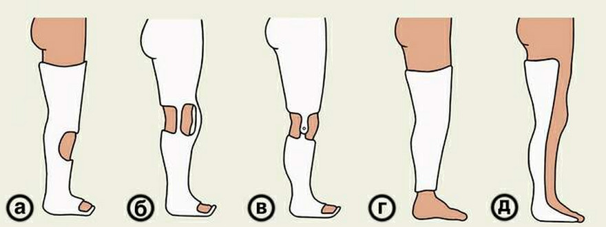 Such an immobilizer can be:
Cartilage disorders are compensated by taking special class drugs.
All this time is divided into such periods:
A compulsory condition for recovery is regular medical support - X-ray checks, periodic examinations and corrective measures. Treatment of intra-articular fracturesIt's more complicated. Damaged and "moving" bones require surgery (osteosynthesis). And such manipulations have to be done in four out of five cases. They try to resort to them if the conservative method did not produce results or a fracture of the knee joint is accompanied by a serious circulatory failure and its further treatment is impossible without surgical intervention.
The operation itself proceeds according to the generally accepted scenario, adjusted for a specific case:
Modern materials allow once again not to injure the tissue, keeping them intact. Such work should be done by professionals who know all the subtleties and are clearly aware of the risks of possible complications. By the way, an intelligent doctor will not hide this fact from the patient, but will tell in detail how to prevent them.
In case of serious skin lesions, the fragments are temporarily fixed with the help of the Ilizarov apparatus. When the wound heals, “twisting” of the fragmented parts of the plates is made. It is impossible to delay with such actions - the broken protrusions of the tibia alone almost always entail other undesirable consequences, such as trauma of the meniscus, "crosses" or lateral ligaments. In terms of recovery rarely delayed by more than 2-3 months. Although the treatment can be continued if complications arise. ComplicationsExperienced doctors know that such injuries are often accompanied by various complications. In the postoperative period, so-called local manifestations may appear - in the form of suppuration or infection. A broken knee joint, more precisely, its injury, can give a “failure” depending on the nature of the shift. A damaged calyx may, in time, respond with arthrosis, in which severe pain is felt in the front.
Other problems may include:
Therefore, competent rehabilitation acquires exceptional importance (regardless of age). Rehabilitation periodWhen selecting a recovery program, specialists try to take into account the complexity of each individual case. But there are general recommendations that should be followed:
The complex of physical therapy will require the patient to be careful and understand the essence of the exercises. Often they are prescribed to begin 2-3 days after surgery. Of course, these are simple exercises, but over time they will become more complex, and the number of repetitions will increase. Conventionally, physical therapy techniques for such problems can be divided into three groups. And if the first two are aimed at overcoming pain and combating atrophy, the third is already a “pure development” of a limb:
Recall that the self-activity for such serious fractures is not suitable, as well as an unauthorized increase in loads. It is also undesirable to take long breaks in classes. We figured out what are fractures of the knee, and how to treat them. We hope that such injuries will bypass you. The most dangerous among the fractures of the lower limb is a fracture of the knee. It is no secret to anyone that the limb for the most part functions at the expense of the knee. Bending and bending it, people can walk, run, jump, sit down. Fracture of the knee joint violates the functionality of the leg, thereby depriving a person of the ability to move. The human knee joint includes:
So with a fracture of the knee, the integrity of any of the bones entering the joint can be broken. VarietiesFracture of the knee can be open or closed. An open fracture is more dangerous, since a fracture of this type causes damage to a large amount of soft tissue and skin. At the same time there is bleeding from damaged vessels, and fragments of bones are visible in the wound. The injury can be complete when the bone breaks into two or more fragments (with a comminuted fracture), or incomplete, when only a crack appears in the bone. There is a condylar fracture when the fault line passes through one or both condyle of the femur or tibial bone, as well as a fracture of the kneecap. These injuries can be combined with dislocation of the leg or with a fracture of the body of the thigh. If the bone fragments have shifted from their physiological location, then this is a fracture with an offset. Such fractures are dangerous in that displaced bone fragments can damage the surrounding tissue and form an open type of injury. This type of damage is treated a little longer than a fracture that is not complicated by the displacement of fragments. The reasonsThe knee condyles most often break during heavy physical exertion on the leg, the cause of the calyx damage is almost always a strong direct blow. Damage of an open type may occur due to a fall from a great height or through a traffic accident. Pathological fractures occur even with a slight force, the factors that lead to them are diseases of the musculoskeletal system (osteomyelitis, osteoporosis), bone tuberculosis, oncology, obesity, diabetes, calcium deficiency, etc. More often, injury occurs in:
For whatever reason, an injury would occur, the victim should immediately be given first aid and taken to the trauma center. Clinical pictureWhen an injury occurs, the signs of it should serve as a reason for going to the doctor. Symptoms of a fracture of the knee joint, or rather the degree of their severity depends on the type of injury. With a crack in the knee, a person may feel a slight pain syndrome that does not interfere with his movement. For complete fractures, the symptoms are as follows:
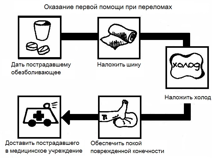
With an open type of injury, a wound and bleeding occurs. After the discovery of symptoms indicating the presence of a fracture, the person must be given first aid and call an ambulance. Providing first aidWith an open fracture, there is a risk of large blood loss, so if a person is injured, the first thing to do is to stop the bleeding. To do this, you need to figure out what is damaged - the artery or vein. The blood from the vein is bright red, flowing evenly and not very strong trickle. Arterial blood has a maroon color and beats almost a fountain. With venous bleeding, the tourniquet does not overlap; it is enough to put a sterile napkin soaked in hydrogen peroxide on the wound and press it with your fingers for a few minutes. If blood flows from an artery, then to prevent large blood loss and traumatic shock, it is worth putting a tourniquet on the lower third of the thigh. The leg should not be tightened longer than forty minutes, so you should write down the exact time of imposition on a sheet of paper and put it under the harness. It is also necessary to monitor the color of the fingers on the injured limb, if they began to turn pale, became cold and numb, it is worth loosening the tourniquet. After the blood stops flowing, it is worth treating the wound edges with an antiseptic and apply a sterile dressing. The next step is to fix the limb in a stationary state by applying the tire. The limb should be fixed in the position that it assumed when it received the injury.
As a tire, two or three long, hard objects can be wound, which can be wound with bandages or with any cloth to the foot. The material for the tire must be sufficiently long, since not only the knee is to be fixed, but also the two nearest joints - hip and ankle.
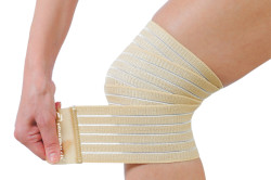
With strong pain, you must give the victim an analgesic drug. An analgesic injection is ideal for anesthesia, but if the person providing first aid does not have the skills, then it is better not to risk taking pills. Anesthetize the place of injury and prevent severe swelling can a cold compress. Ice or a frozen product must first be wrapped in a cloth in order not to freeze the skin. Do not apply a compress directly to the wound, if it is present. After rendering first aid, a team of medical workers should be called, who, if necessary, will apply the tire correctly, make additional anesthesia and, putting the injured on hard stretchers, will take him to the Department of Traumatology for examination, diagnosis and treatment. DiagnosticsWhen a condyle condom fracture occurs, treatment does not begin until the doctor has performed the examination and has made an accurate diagnosis. First, the doctor finds out what caused the injury, will make an external examination of the injury site, will palpate. After that, the patient is taken to an X-ray. In the picture, the doctor sees the location of the fracture, determines its type and number of fragments. However, the cartilage tissue is not visible on the X-ray, therefore arthroscopy is additionally done to the patient. Using an arthroscope, you can see the condition of the cartilage and meniscus of the knee, as well as diagnose hemarthrosis. In severe cases, magnetic resonance or computed tomography. It is mandatory to take the general analyzes of urine and blood, as well as blood for hemoglobin with a strong blood loss. Medical eventsFor fractures without bias, treatment is carried out by a conservative method. Local anesthesia is administered to the patient, the limb is straightened, and a plaster bandage is applied to it, which captures the lower leg and half of the thigh. Additionally, the patient is prescribed analgesics, drugs against edema and inflammation. With a simple fracture, the bone grows together in a month and a half. At the time of treatment, the patient must move using crutches. Restoration of joint mobility and muscle strength after conservative treatment requires great effort.
After all the manipulations, the doctor stitches the edges of the wound and applies plaster for a month and a half, leaving a window over the wound for its subsequent processing. After the operation, the patient should keep the limb in a raised position, placing a cushion under the ankle, and wiggling with the fingers so that no thrombosis occurs. On the second day after surgery, the patient is allowed to stand up and even lean on the injured limb, but with such force that it does not damage the plaster cast and prevent painful sensations. Periodically, the patient is given an x-ray to track the dynamics of treatment and bone splicing. Recovery periodRegardless of the way in which the treatment was carried out, rehabilitation after a fracture of the knee is a mandatory step on the road to recovery. The attending doctor prescribes physical therapy, massage, physiotherapy, and a special diet. The patient should consume foods rich in calcium, and exclude from his diet pastry, coffee, strong tea and alcoholic beverages. The patient should walk as much as possible in the fresh air, especially in sunny weather. The ultraviolet rays of the sun fill the body with vitamin D, without which the calcium supplied with food simply cannot be absorbed. With a competent approach to rehabilitation and self-reliance, a person relatively quickly returns to an active lifestyle, and the injury leaves no consequences. |
 Clinical cases of fracture of the knee are divided into many types. The main ones are. The second large group is the division of this type of injury on an extra-articular fracture and intra-articular. To aggravate the diagnosis can displace bones of a different nature.
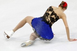
 In some cases (if the bones have greatly shifted or split into numerous fragments) X-rays are not enough, and computed tomography may be prescribed as an additional examination.
 An ambulance doctor will examine the injury and be hospitalized in the hospital, where the patient will have a more detailed diagnostic examination. It includes a primary survey and a visual inspection of the damage. Next, an x-ray will be assigned, perhaps not one, but from several angles to see the severity of the injury. In some cases, X-rays are insufficient and CT scans are indicated.
 Everyone can determine open fractures - the skin will be damaged, which is accompanied by bleeding of different intensity. Recognizing and accurately identifying closed damage is more difficult, in such situations it is better to provide all possible assistance and to call a doctor.
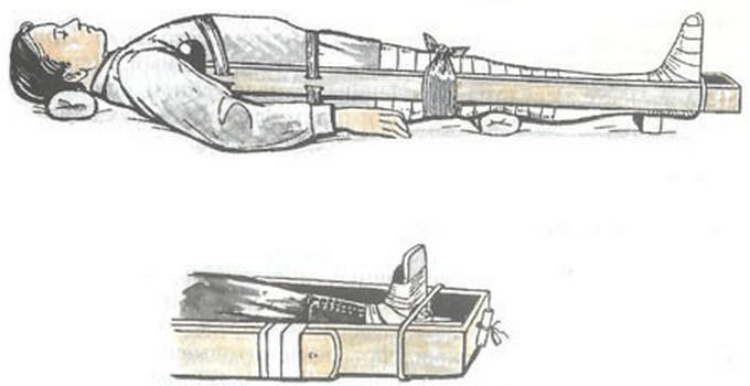 Do you know? 100 years ago, bed rest was considered almost a panacea. And only at the turn of the 1910s of the 1920s. The benefits of physical therapy have been proven.
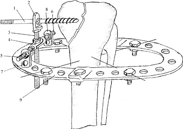 Did you know? In a state of traumatic shock, a person may not feel pain at all. Sensitivity returns, some time after realizing what happened.
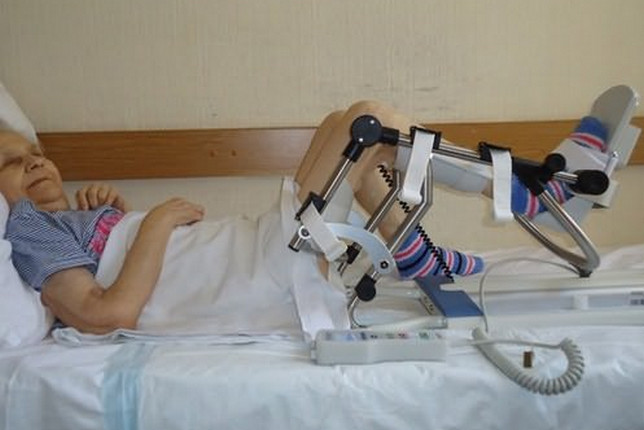
| Read: |
|---|
Popular:
How to moisturize dry skin in winter
|
New
- Owl tattoo and its meaning for girls and guys
- Chicken with honey and mustard
- Is a single kidney pregnancy possible?
- Varnish for stone with a wet effect
- How an optometrist checks eyesight
- How to take grape oil
- Getting a driver's license: restrictions on vision
- Skin care at home
- Inexpensive personal care products
- How to get margarine and how to choose it correctly?


