Site sections
Editor's Choice:
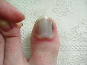
- Manifestations and recovery in case of marginal fracture of the first toe
- Wine fermentation at home
- Chicken with mushrooms - recipes with photos
- Treatment and rehabilitation for knee fracture
- Chicken fillet with porcini mushrooms in a pan
- What drugs lower cholesterol
- How to accelerate the healing of a leg fracture
- Plaque formation and composition
- How hair straighteners work
- Technological map of the lesson the structure of seeds of dicotyledonous plants
Advertising
| Open fracture of the finger. Manifestations and recovery in case of marginal fracture of the first toe |
|
Violation of the integrity of the phalanges can be complete or partial. Traumatic and pathological are distinguished by origin. The cause of the pathological disorder is various kinds of diseases that make the bones less durable. These include tumors, osteoporosis, osteomyelitis, hyperparathyroidism, tuberculosis, etc. Traumatic are much more common. They are received during incidents, accidents, running, accidental strikes, playing sports, falling objects, etc. A marginal fracture of the toe means that a thin flat fragment is separated from the bone. This is not a very serious injury, which most often occurs when running. A fracture can affect one phalanx:
Or a few, then they are called matching. The thumb comes forward and carries much stronger loads than the rest, so it is he who most often suffers. How to determine?With a violation of the integrity of the phalanx bone, the following symptoms appear:
To determine a fracture of the thumb, an x-ray is necessary. The doctor also examines and interviews the victim. Fractures of the thumb are very painful, so the patient is immediately injected with anesthetic medicine. The main task for splicing is to fix the fragment and immobilize the finger so that the bones can grow together. Incorrect splicing will result in the operation having to be performed. With minor damage, the patient is prescribed rest, ice, compresses and recovery. Rest is necessary to completely remove the load from the leg, ice serves as an anesthetic, compresses relieve inflammation and swelling, and lifting reduces blood flow so that it does not accumulate in the limb. At first, cold compresses should be applied every hour for 10-15 minutes. This will help relieve acute pain and swelling. It is also recommended that you apply bandages from an elastic bandage from time to time if you walk or move your leg. But they cannot be kept for long, otherwise limited bleeding will lead to even greater problems. As a rule, mild fractures heal in 4-6 weeks, after which you can return to your usual lifestyle. Recovery period
For faster recovery, therapeutic massage sessions, physical procedures, and special gymnastics are carried out strictly according to the doctor’s instructions. The diet should include a maximum of foods with protein and calcium. PreventionAlthough such injuries are not very scary, it is better to avoid them. You can be cautious and prevent such damage:
At the slightest sign of a fracture of your fingers, take pain medication, try to fix the foot and consult a doctor immediately. Previous NextFracture localization: Nail; average; combined; main. Doctors also classify fractures by how the bone was destroyed: With the formation of fragments; a fracture in which bone fragments are displaced; fracture without bone displacement. The reasonsThe main causes of the formation of a fracture are ailments that contribute to the fact that the bones become not so strong. This occurs when: Tumor osteoporosis; osteomyelitis; hyperparathyroidism; tuberculosis. Plaster cast application Also important reasons for which a fracture can be obtained are injuries sustained during accidents, playing sports, or hitting heavy objects. 0 0 Closed fractures of the phalanges and metacarpals account for 31.2% of closed fractures of the bones of the hand, fractures of the distal phalanges - 18.2%, medium - 37.5%, proximal - 21.4%, metacarpal bones - 13.3%, sesamoid bones - 0.6%, wrist bones - 9%. Intra-articular fractures in the distal interphalangeal joints The most characteristic and frequent type of fracture is the separation of the back edge of the base at the place of attachment of the extensor finger. The size of the bone fragment and the degree of displacement are essential for the outcome (Fig. 112). Fig. 112. Options for separation of the back edge of the distal phalanx. We distinguish four degrees of damage (E. V. Usoltseva, 1939). First degree: a small fragment of the bone, the articular surface was not affected. Displacement and deformation are negligible; distal ... 0 0 Intra-articular fracture - damage to the bone inside the joint capsule. This is a complex trauma that requires increased attention from the traumatologist and a special approach to treatment, since the range of movements depends on how accurately the articular surfaces have been restored. It can occur in the area of any joints, both large and small. Due to possible long-term consequences, intra-articular fractures of large joints of the limbs are of the greatest clinical significance. The joint consists of two or more matching ends in the shape of the ends of the bones, which are interconnected by means of a capsule and ligaments. The capsule forms a cavity isolated from other anatomical formations. Ligaments are located inside and outside the capsule that hold the bones, preventing them from moving too much. The ends of the bones are covered with smooth cartilage, thanks to which the bones can glide freely relative to each other. A small amount of fluid is contained inside the joint –... 0 0 An intra-articular fracture is called damage in which the bones are destroyed, and the fracture line is located in the joint cavity (partially or completely). These injuries have some features that distinguish them from the category of similar injuries, and require a more thorough approach to diagnosis and rehabilitation. The peculiarity of these fractures is that in addition to the damaged bone, the joints are also involved in the process, in violation of the consistency of the surfaces of the joints with possible shortening of the tendons and their poor mobility in the future. AnatomyJoints are a complex part of the musculoskeletal system with articulated properties, due to which the movement of the limbs is ensured. Their normal functioning is disrupted as a result of altered anatomical relationships of the elements of the joints that arose after the injury. The most serious consequences arise with fractures of the intraarticular departments that perform support and ... 0 0 Among all bone fractures, the data is 5%. Fractures of the second finger are more common, in second place is the fifth finger. In almost 20% of cases, multiple fractures of the phalanges of various fingers are observed. More often damage to the main phalanges occurs, then the nail phalanges and rarely - the middle phalanges. AnatomyFour of the five fingers consist of three phalanges - the proximal (upper) phalanx middle and distal (lower).The thumb is formed by the proximal and distal phalanx. The distal phalanges are the shortest, the proximal phalanges are the longest. Each phalanx has a body, as well as a proximal and distal end. To articulate with adjacent bones, the phalanges have articular surfaces (cartilage). The reasonsFractures occur at the level of the diaphysis, metaphysis and pineal gland.They come in without bias or with bias, open and closed. Observations show that almost half of phalangeal fractures are intraarticular. 0 0 Fracture of a finger on a hand how much to wear gypsum Home / Diseases of a finger on a hand how much to wear a gypsum Finger fractureNo one is immune from injuries of varying severity, especially brushes, because when you fall, a person tries to avoid injury with the help of these parts of the body. Finger damage can also occur due to improper handling of mechanisms that exert a twisting or squeezing effect. The nature of the fracture, and therefore its treatment, also depends on how the finger is damaged. Finger fracture causesMost often, fingers are injured in athletes: volleyball players, basketball players, gymnasts and boxers, although the latter are protected with special gloves. Also at risk are children and people with osteoporosis. In any case, a fracture is always due to direct injury: Hit; falling heavy object on the brush; getting the brush under the moving mechanism. Fracture of the finger: the main ...0 0 A fracture of the finger is a fairly common injury. In the human skeleton there is a huge number of tubular bones, but the smallest of them are represented by the phalanges of the fingers. ClassificationDepending on the location of the line of fracture of the phalanx of the finger, fractures are divided into: Diaphyseal part; Periarticular; Intra-articular fractures of the proximal end; Intra-articular fractures of the distal end. By the type of fracture line: Transverse; Helical; Slanting; Regional; Comminuted. With some injuries, a person experiences a fracture of several phalanges or fingers. If the victim has a fracture of the middle phalanx, then very often there is a displacement of bone fragments at an angle open to the back of the hand. Damage to the terminal phalanx, as a rule, occurs under the influence of a direct blow to the fingers of the hand. With this type of fracture, several small fragments and a bruising hematoma with exfoliation of the nail are formed ... 0 0 The brush, as an important functional organ in human life, is very often injured. Fractures of the bones of the hand account for almost 1/3 of all fractures of the skeleton. Damage most often occurs in people of working age, mainly in men. Fractures and fractures of the wrist bones are rare; make up about 1% of fractures of the bones of the hand. The scaphoid is more often damaged, less often - lunate, trihedral. Fractures of other bones of the wrist are extremely rare. As a rule, all of them are intraarticular. Scaphoid fractureA scaphoid fracture occurs more often as a result of a fall on the hand. With a radial deviation of the hand at the time of the fall, the process of the radial bone acts on the scaphoid as a splitting wedge. The fracture line then coincides with the gap of the wrist joint, and during movement one fragment of the scaphoid remains with the proximal row of bones, the second moves with the distal. Therefore, this fracture requires ... 0 0 Fractures of the fingers are often found in medical practice. One of the most common injuries is considered a fracture of the big toe. It occurs when heavy objects fall on the foot, hit on the finger, and when the foot is twisted. The big toe is anatomically and functionally different from other toes of the lower limb. The clinical picture, treatment and rehabilitation of trauma has its own characteristics. You can read about the features of the fracture of other toes here. The reasonsA fracture of the first toe occurs when a force is exerted on it that exceeds the strength of the bone tissue. Bones are most vulnerable in childhood and old age. In children, the skeleton is in a period of active growth, as a result of this, the bones do not have sufficient strength. Due to age-related changes, the bone tissue of older people does not contain enough calcium, which makes bones fragile and increases the risk of fractures. Big toe injuries ... 0 0 10 CONSEQUENCES OF HIP FRACTURE. A hip fracture is dangerous due to the development of severe traumatic shock and significant blood loss. With medial fractures, a hematoma can form in the trochanteric or inguinal regions. Later complications may develop: prolonged fracture failure and the false joint of the femoral neck, after open intraarticular osteosynthesis, aseptic necrosis of the femoral head, deforming arthrosis of the hip joint, anaerobic infection may join in with open fractures and osteomyelitis may develop. Due to a violation of the intraosseous arterial network, blood supply suffers, which negatively affects bone regeneration processes, and avascular necrosis of fragments develops. With lateral fractures of the femoral neck, a complication may develop in the form of impaired adhesion of the fragments - rocking of the fracture and its incorrect fusion with deformation and shortening of the limb. With a fracture of the distal end of the femur, the knee ... 0 0 11 Features of intraarticular fracturesJoints are complex structures of the musculoskeletal system. Their inherent functional features include providing static loads and kinematics of the limbs, as the joints have hinge properties. In this regard, a dysfunction of the joints occurs with any damage accompanied by a change in the anatomical relationships of the joint structures. The most severe consequences regarding support and kinematic functions are caused by a violation of the integrity of the meta- and epiphyseal (intra-articular) departments of the articulating bones. Violation of the congruency of the articular surfaces observed during such fractures, even after specialized treatment, can lead to the development of degenerative changes, persistent pain syndrome and restriction of movements. In order to predict post-traumatic disorders, fractures with insignificant and significant displacement of fragments, as well as stable and ... 0 0 12 Fracture of a finger of the hand is a common pathology that traumatologists regularly encounter. The short tubular bones that make up the phalanges of the fingers have a rather fragile structure and are prone to injury. Fortunately, pathology is easy to suspect and easy to treat. The main thing is not to delay referring to a specialist, so that a finger fracture is not complicated by severe concomitant conditions. The reasonsThe human brush consists of three departments. Closer to the forearm is the wrist formed by eight small spongy bones. The middle section of the hand is metacarpal bones in an amount of 5, which are directed from the wrist to the proximal phalanges of the fingers. Finally, the fingers themselves are made up of bones called phalanges. Each finger, except the thumb, has 3 phalanges: proximal (closer to the base of the palm), middle and distal (nail). The thumb consists of two phalanges. All damage is divided by 2 ... 0 0 13 The human body is a complex system that requires attention and care. Every day we are faced with various situations in which, under external influences or physical exertion under certain conditions, it is likely to get damaged. Bone fracture is one of the most popular injuries when, before complete healing, a long treatment and a long period of rehabilitation may be required. Most often, fractures of the limbs, as they are more vulnerable. Next, we will consider in detail a toe fracture, signs, characteristic symptoms, methods for diagnosing and treating trauma, as a common type of bone and joint fracture. Briefly about the anatomy of the footFoot is a part of the musculoskeletal system of a person, the most remote part of the lower extremity from the body, which is responsible for a stable position, balance, and movement of the body in space. The foot has a flexible, flexible and movable vaulted structure, which ... 0 0 14 How to accurately identify a fractured finger and make the treatment of injury the most effective Fingers are our main helpers for performing movements in everyday life and at work. A broken finger left untreated or improperly cured can cause many inconveniences to its owner. Since a person uses his fingers in all areas of his life, to describe all the situations in which he can injure them is an extremely long task. Anatomy and functionsThe fingers are located on the limbs and serve to perform tactile and manipulative functions. Fingers consist of short tubular bones connected by tendons. Muscles are located on the bones for their mobility, as well as small and medium vessels. Classification of fractures and their characteristicsFractures are divided into traumatic and pathological. A traumatic fracture is damage to a finger bone ... 0 0 15 Finger phalanges fractures They arise from direct injury and are more often open. Fractures can be periarticular, intraarticular and diaphyseal. Fractures of the nail phalanx are usually comminuted. Clinic. Pain, swelling and limitation of finger function. In the presence of displacement, deformation is noticeable. In fractures without displacement, signs of damage can fit into the picture of a sprain or bruise. Therefore, an X-ray examination is mandatory even if a fracture is suspected. Treatment. In case of fractures of the main phalanx without displacement, the finger is immobilized with a gypsum or aluminum splint when the main phalanx is bent by 45-50 °, the middle by 60 °, and the nail to 15 ° for 3 weeks. Then prescribed therapeutic exercises with the use of physiotherapy. Disability is restored after 4 weeks. In fractures of the main phalanx with displacement under local anesthesia, a closed comparison of fragments is carried out by traction along the axis, pressure on ... 0 0 16 Varieties, signs and treatment of intraarticular fractures In our life, very often there are various injuries associated with an active lifestyle at a young age and various diseases (the first of which is osteoporosis) in the elderly. In addition, accidents and accidents that are not uncommon in the modern world lead to injuries of varying severity. A serious bone skeleton injury is a fracture. Among such injuries, an intraarticular fracture is especially distinguished, because in connection with the anatomical features it requires a more careful attitude, a thorough diagnosis and high-quality treatment and rehabilitation. Concept and varietiesThis type of injury is characterized by damage to that part of the bone that is near the joint and takes part in its formation. As a result, there is a violation of the function of the bone joint, limitation of mobility, damage to the intraarticular capsule, which requires a longer ... 0 0 17 A bone fracture is a pathological condition in which there is a partial or complete violation of the integrity of its anatomical structure under the influence of an external force. Fractures of the forearm can develop due to mechanical injuries (when falling on the arm, hitting the forearm, falling something heavy on the arm, etc.) or occur as a result of certain diseases (osteoporosis, rickets, osteomyelitis, bone tumor, etc.), accompanied by a violation of the incorporation of minerals into bone tissue. Fractures of the forearm are a fairly common pathology, characterized by a wide variety of clinical symptoms. With such fractures, pain, swelling at the site of damage, external bleeding, bruising, impaired skin sensitivity, deformation of the forearm, impaired function of the elbow and wrist joints with the restriction of active and passive movements can occur. With open fractures in the wound, bone fragments can often be seen. With fractures of the forearm are possible ... 0 0 18 Fractures of the phalanges of the fingers are common injuries. Unfortunately, despite the high incidence of these fractures, treatment outcomes are unsatisfactory. This is explained not so much by the severity as by underestimating the severity of the damage, errors in the diagnosis and treatment of phalangeal fractures. It must also be emphasized that the brush, which is the thinnest tool that produces a variety of movements that cannot be repeated by any artificial mechanism, is often injured. The restoration of the function of a damaged hand is, first of all, the restoration of anatomical ratios, without which the renewal of finger movements is impossible. The mechanism of injuries is most often direct (a blow to the fingers, the fall of heavy objects on the hand). Fractures of the phalanges, especially the main one, can also occur with indirect injuries - a sharp over extension, twisting of the finger. The nail is most often injured, less often the main and even less often the average phalanx. More often... 0 0 19 Types of deformation of the radius Fracture classification The main causes of radial fractures First aid for suspected fracture Treatment of a radial fracture Hand development after fusion, rehabilitation Warm-up Training What does the radius bone look like? All fractures associated with radial bones are divided into two types according to the nature of bone displacement: Flexion, Extensor. Fracture classificationIntra-articular ....0 0 20 Damage to bones can be severe, in which not only a bone crack is formed, but several fragments. This condition is called a comminuted fracture and is most often accompanied by the displacement of small fragments of a broken bone. The treatment of this form of fractures is associated with certain difficulties and depends on the location (location) of the fracture, the form of damage, the number of fragments, their shape and size. Various factors can serve as the causes of the occurrence, but most often the impact of blunt objects on the limbs, a fall, sports injuries, unsuccessful or careless movements causes a fracture. One of the reasons that not only contribute to the occurrence of fractures, but can also lead to complications, are metabolic disorders and diseases associated with these conditions, which lead to increased fragility of bone tissue. As a result of such violations, instead of the usual fracture, comminuted occurs, since fragile tissue is destroyed not only when ... 0 0 22 Making a broken finger is easy, getting injured in everyday life or playing sports. Often this happens during a fall, as instinctively a person tries to defend himself, exposing his arms as a support. Signs and effects of fractureThe following signs indicate a fracture of a finger: it is a aching pain, swelling of tissues, restriction of movement, deformation of the appearance of a finger. In case of a fracture with an offset, a bluish tint is also observed at the site of injury. Often the injury is accompanied by a bruise of the nail plate. If there are signs of a fracture of the phalanx of the finger, it is necessary to contact the nearest emergency room, since in order to make the diagnosis more accurate, it is necessary to take an X-ray. To preserve the function of the hand in full after injury, it is important that the doctor deals with the treatment of each case. To treat the fracture of the phalanx as something insignificant is a serious mistake that can lead to unpleasant consequences: ...0 0 23 Fracture is a violation of the linear integrity of the bone under the action of a force that exceeds the tensile strength of the bone. The main cause of fractures in the world is injury. In disease statistics, he ranked third. Fracture is a serious pathology, despite the small size of this part of the body. According to statistics, finger fractures account for 5% of all fractures. Fractures of the fingers are severe injuries of the hand, since they significantly reduce its functionality. Diagnosis of a fracture of a finger, as a rule, does not cause difficulties, but with the treatment the situation is different. To fully restore the shape and function of the bone, it is necessary to strictly follow all the recommendations for the treatment of this pathology. Deviation from treatment requirements leads to serious complications and even disability. Anatomy of the handHuman hand is extremely difficult from the point of view of evolution, education. It consists of 30 - 32 ...0 0 24 What to do with a broken finger? Hello Yulia. A phalanx of a broken finger is not likely to bend because you are not developing it. Fractures of the fingers on the localization is divided into extra-articular and intra-articular. A traumatic fracture is damage to the bone of a finger due to injury. Our fingers perform very subtle, coordinated movements and the violation of these movements can have a huge impact on the daily and professional activities. To preserve the function of the hand in full, it is very important that all fractures of the fingers be evaluated by a doctor to determine the appropriate treatment. If you think that a broken finger is a minor injury, then you are seriously mistaken. If you have symptoms of fracture of the phalanx of the finger, you must contact the emergency room at the place of residence. The doctor must determine not only the location of the fracture, but also the type. If a fracture affects a joint (intra-articular fracture), it is important to ensure that the articular surface does not ... 0 0 25 Fracture of the finger on the hand - an injury that occurs quite often in everyday life. Such damage can permanently knock a person out of the usual rut, since it becomes impossible to perform even the simplest work. If you have a broken finger, you should immediately seek medical help. Otherwise, the bones may grow together incorrectly, and the hand will cease to function. This is especially true for cases of a broken thumb, since it accounts for the main burden when performing any manipulations. Hand anatomyIn the process of evolution, the mammalian brush underwent many changes that helped to adapt to the constantly changing habitat conditions. Thanks to its complex organization, the human brush is extremely functional. It consists of 27 bones. Each finger consists of three small stones, called phalanges. The exception is the thumb, in the composition ... 0 0 Fractal phalangeal fractures refer to common damage. Unfortunately, despite the high incidence of these fractures, treatment outcomes are unsatisfactory. This is due not so much to the severity as to the underestimation of the severity of the injury, to errors in the diagnosis and treatment of phalangeal fractures. It should also be emphasized that a brush, which is a very fine instrument that produces a variety of movements that no artificial mechanism can repeat, is often subject to injury. The restoration of the function of a damaged hand is, first of all, the restoration of anatomical ratios, without which the renewal of finger movements is impossible. Injury mechanism most often direct (hit on the fingers, the fall of heavy objects on the brush). Fractures of the phalanges, especially the main one, can also occur in the case of an indirect injury - a sharp over-flexion, twisting of the finger. The nail is most often injured, the main phalanx is more rare and even less often. II finger is most often damaged, followed by III, and then the rest. Quite often several fingers are injured at once. Diagnosis of phalangeal fractures does not cause much difficulty. First, for these fractures characteristic mechanism of injury. Secondly, due to the fact that from the back of the phalanx are covered with a thin layer of soft tissue, despite the swelling, the phalanx deformity is clearly visible. Some difficulties arise in the recognition of fractures without displacement, cracks and intra-articular fractures. However, in these cases, the mechanism of injury makes it possible to suspect damage to the hand. In cases of phalangeal fractures, the usual signs of bone fractures are observed: swelling and hemorrhage in the area of the fracture, the presence of deformities, pain on palpation of the area of the fracture, and impaired function of the injured finger. All these signs are present at the displacement of fragments. Severe deformity is absent in non-displaced fractures (Fig. 47, 48), fractures and intra-articular lesions. In transverse fractures of the main phalanx, a characteristic displacement of fragments is observed - at an angle open to the back side, due primarily to the direction of the traumatic force, as well as the traction of the womb-shaped and interosseous muscles. The proximal fragment is displaced to the palmar side, and the distal fragment is displaced to the rear side, resulting in an angle open to the rear side. Fig. 47. Intra-articular fracture of the middle phalanx of the finger. The nature of the displacement of fragments of the middle phalanx depends on the level of the fracture of the phalanx and the location of the line of fracture in relation to the place of attachment of the tendons of the surface flexor of the finger. At the fracture of this phalanx distal to the attachment of the tendon legs, the proximal fragment under the action of thrust is displaced to the velor side and an angle open to the rear side between the fragments. In case of a fracture proximal to the attachment of the legs of the tendon, the distal fragment will tilt in the palmar direction, forming an angle open to the velor side. Permanent and characteristic is the displacement of the fragment with tear-off fractures on the back of the nail phalanx. These fractures occur when an indirect injury - a sudden sudden bending of the nail phalanx (hitting the nail phalanx on solid ground, falling on a slightly bent finger, hitting the tip of the finger with a volleyball, etc.). In these types of injuries, a rather strong tendon of the extensor of the nail phalanx is strained, which leads to the separation of a piece of bone from the dorsum of the nail phalanx. Usually this is a piece of the base of the nail phalanx. In this case, the nail phalanx hangs and the patient cannot actively unbend it. X-ray in two projections allows to clarify the presence and nature of the displacement of fragments.
Fig. 48. Fracture of the main phalanx of the second finger (a) and double fracture (b). Treatment of phalangeal fractures is reduced to a comparison? displaced fragments, fixing them for the time required for consolidation, and the subsequent restoration of function. Each of these stages of treatment is important; neglect of one of them can lead to serious consequences. Before setting out the method of treating phalangeal finger fractures, it is necessary to recall some of their anatomical features. The lateral ligaments, which strengthen the interphalangeal joint, are located somewhat eccentric with respect to the heads of the phalanges. Due to this, with a straightened finger, they are in a relaxed state, with a bent - stretched. When a phalanx fracture is injured, not only the bone, but also the soft tissues surrounding it (tendon sheath, fascia), hemorrhage into the joint capsule, lateral ligaments and walls of tendon sheaths occurs. If the finger is left in a position unfavorable to the function for a long time, then due to scarring of the soft tissues, subsequent recovery of the function may be difficult. For example, if you keep your finger extended for a long time, the lateral ligaments of the interphalangeal joints shrink and do not allow the finger to bend after the fusion of the fragments. The first condition in the treatment of phalangeal fractures: the finger must be given a functionally advantageous position, the second: only the damaged finger is fixed, the rest must remain free, and finally, the third condition: the fixation should be short-lived, since along with the consolidation of fragments, fusion between the bone and the damaged tendon can occur the vagina, which also makes it difficult to restore movements of the damaged finger. Comparison of fragments should be carried out necessarily under anesthesia. Soft tissue tight bone. The introduction of novocaine creates even greater tension in already strained tissues, which is not always safe; therefore, it is best to apply conduction anesthesia according to Obegu-Lukashevich. In the interdigital spaces on both sides of the damaged finger injected with 10 ml of 1% solution of novocaine. At the same time, sufficient anesthesia of the injured finger is achieved, allowing it to be painlessly repulsed from breaking. In each trauma clinic or clinic, there must be a set of wire tires for fingers (fig. 49). Before starting the reposition, prepare the tire, cover it with a strip of thin cardboard cut out of its shape, cover the tire with a thin layer of cotton wool and wrap it with a bandage.
Having thus prepared the structure, a circular plaster bandage is applied from the middle third of the forearm to the metacarpophalangeal joints and the splint is inserted so that it is located along the palmar surface of the injured finger. When screwing in, the tire should be set so that its free end protrudes 1-2 cm above the fingertip. Having laid the finger in this way, strengthen it with several rounds of adhesive tape, starting just below the nail. Then go to the X-ray room, where, under control, the tire is bent over the fracture site. At the same time, pinned finger, following the bend, pulls the broken phalanx and fragments are placed in the correct position. Care must be taken to avoid hypercorrection. When the tire is bent, the finger takes a half-bent position, which is favorable for the function (fig. 50). It is necessary to emphasize once again that accurate matching of fragments is crucial for the restoration of function. Thus, fragments are mapped at an offset at an angle open to the rear.
When displaced at an angle open to the palm side, which occurs when the middle phalanx fractures, after the tire has been inserted, it is bent so that the finger laid on it takes a half-bent position. Then, in the x-ray room, pressing the top of the angle formed by the fragments, the fragments are repaired. Checking the correct position of the fragments, the finger is fixed to the tavern several rounds of adhesive tape. Active movements with intact fingers must be made from the first days after the injury. They help to improve the blood supply to the hand and reduce swelling. The patient should be explained the importance of active movements with intact fingers. Fixing a broken finger lasts 2-3 weeks. After X-ray control, immobilization is removed and active movements are recommended. With accurate matching of fragments and good adhesion, finger function and working capacity are restored in 3-4 weeks. The physiotherapy room must have a set of devices to restore function. However, the best way to restore the function of damaged fingers are active movements that the patient produces throughout the day. In case of phalanx fractures without displacement of fragments, they should also be fixed with the help of a wire bar in the functionally favorable position of the damaged finger for a period of 10-12 days. The ability to work is restored after 2-3 weeks. The method described above for the treatment of phalangeal fractures of the fingers in most cases makes it possible to compare and fix fragments for the period necessary for good fusion. However, there are cases when the break-ups are poorly matched and tend to re-shift, especially with oblique fractures. In such cases, surgical intervention and fixation of fragments by intraosseous injection of the spokes are shown. Another type of fixation has to be applied when tearing off a piece of bone from the rear of the nail phalanx. On the finger impose a circular plaster cast. In order that it does not slide off the finger, it is fixed on the brush with several rounds of a naked gypsum bandage. Then, while the gypsum is not hardened, the nail phalanx of the plastered finger rests on the table, creating its over-bending. With this injury, the detached bone piece does not move far away, therefore, when re-bending, the nail phalanx approaches it. In the position of re-bending the finger is 3-4 weeks. After removing the bandage, the finger function is quickly restored. In case of long-term fractures, as well as in cases where the fragment is not matched with the nail phalanx or, hurting at the joint, it causes pain, surgical removal of the fragment and stitching of the ruptured extensor tendon and the capsule are shown. Very often with fractures of the nail phalanx, subungual hematoma is formed, which causes no less pain than a fracture. In these cases, before applying the fixing splint in the Foot, a hole should be made and the hematoma removed. Dubrov Ya.G. Outpatient Traumatology, 1986 |
 About six weeks after the fracture, it is important not to overexert or injure the limb again. This is easy to do - limit active movements, do not play sports and do not take long walks.
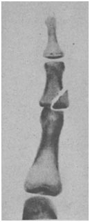
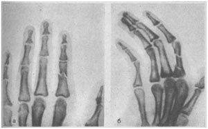
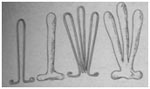 Figure 49. Wires for treating phalangeal fractures.
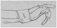 Fig. 50. Fixing a finger on a wire bar mounted on a plaster splint.
| Read: |
|---|
Popular:
The most dangerous poison for humans
|
New
- Amanita mushroom - medicinal properties, how to take tinctures and prepare ointment
- Injured with pelvic injuries
- Devices for nail extension: a list of all necessary
- Owl tattoo and its meaning for girls and guys
- Chicken with honey and mustard
- Is a single kidney pregnancy possible?
- Varnish for stone with a wet effect
- How an optometrist checks eyesight
- How to take grape oil
- Getting a driver's license: restrictions on vision


