Site sections
Editor's Choice:
- Symptoms, course, treatment and prevention
- Lecture Topic: Digestive System
- How many grams of honey in a teaspoon and a tablespoon
- Pancreatitis tests: what studies should be done and what do the indicators say
- How to remove cholesterol from the body
- Hyperplasia of the cervix and endometrium: why treatment is necessary
- How much fracture is fused
- How do leaves change in autumn
- All you need to know about pumping honey
- Manifestations and recovery in case of marginal fracture of the first toe
Advertising
| Fracture. Symptoms, course, treatment and prevention. Medical reference book. Medical Encyclopedia. Bone fractures |
|
Bone fracture(fractura ossis) is called a violation of its integrity under the influence of external violence. Pathogenesis (what is happening?) During bone fracturesFractures may be traumatic and pathological (non-traumatic). Pathological fractures occur in acute and chronic osteomyelitis, osteogenesis imperfecta, hyperparathyroid osteodystrophy, bone cysts, benign and malignant bone tumors, bone metastases (more often than others, tumors of the mammary glands, kidneys, prostate, lungs, stomach, etc. metastasize to the bone). . Fractures open and closed. The skin over the fracture and other soft tissues with an open fracture can be damaged by the traumatic force that breaks the bone - these are the first-discovered fractures; if soft tissue and skin are perforated from the inside with the sharp end of the fragment, this is a second-open fracture. With a secondary open fracture, the wound is usually small, equal to the diameter of the end of the fragment perforating the bone. Both with a primary open and a secondary open fracture, primary microbial contamination of the fracture zone takes place, therefore suppuration and osteomyelitis are possible with both fractures. Closed fractures may be complete and incomplete. With incomplete fractures, the integrity of the entire bone is not broken. These are marginal fractures, detachments of tubercles of bones. Localization distinguishes fractures diaphyseal, metaphysical and epiphyseal. Violations of bone integrity in children and adolescents in the germination zone are called epiphysiolysis. Epiphyseal fractures are usually intraarticular. Metaphysical fractures are also called periarticular fractures. Depending on the height of the location, fractures in the lower third of the bone, middle third and upper third are distinguished. It is necessary to distinguish between the concepts of "fragment" and "fragment". The fragment contains all the components of the bone, i.e., if it is a fragment during a diaphyseal fracture, then it certainly has a bone canal. There are two fragments (with a single fracture), three (with a double fracture), four (with a triple fracture). Multi-fragmented as well as large-fragmented fractures are distinguished. Fractures in the direction of the fracture plane transverse, oblique, helical, longitudinal. Fractures occur without displacement of fragments and with displacement of fragments. Displacement can be primary: occurs at the time of a fracture under the influence of a traumatic force and secondary: occurs under the influence of muscle contraction (retraction); on radiographs we usually see a secondary displacement of fragments. Distinguish displacements of fragments along the length, width, at an angle to the axis and rotational. The angular displacement of the bone in a segment with two long bones (forearm, lower leg) is also called axial displacement. Unlike angular, it is difficult to remove with a closed reposition. From a clinical point of view, it is important to distinguish between fractures stable and unstable. Stable fractures have a transverse fracture line. In unstable fractures (oblique, helical) due to increasing muscle retraction after injury, a secondary displacement necessarily occurs. Fractures can be hammered when the end face or the sharp edge of the end face of one fragment is embedded in the end face of another fragment. A classic example of an implanted fracture is an abduction fracture of the femoral neck. With such a fracture, the retraction of the muscles surrounding the hip joint enhances the adhesion (sticking) of the fragments, eliminates the possibility of their displacement. Impacted fractures are also found in fractures of the trochanteric region. In the elderly and senile age, this incision should not be eliminated, since it contributes to a more rapid healing of the fracture. The damaging force during the fracture determines the nature of the fracture and the direction of the fracture line. From an X-ray, seeing the direction of the fracture line, the shape of the fragments, one can imagine the mechanogenesis of the fracture. A fracture from flexion is characterized by a rupture of the bone on the side of its extension (side opposite to the direction of the forces that bend and break the bone) and puncture of a triangular fragment on the side of compression of the bone. A fracture from a shift occurs when one bone abuts against another, under the influence of amplifying external forces exceeding the strength of the bone, breaks, moves it. Fractures from a shift occur in the ankle joint:
The tear-off fracture is opposite to the shift, when a section of the bone is opened by sharply contracted muscles or ligaments with excessive movement in the joint. This is a tear fracture of the medial ankle with the pronational mechanism of injury - the deltoid ligament tears off the inner ankle; the fracture line is always horizontal and often at the level of the joint space. This is a tear fracture of the lateral ankle with the supination mechanism of injury, when the heel-ligamentous ligament tears off the apex of the outer ankle; the fracture line is horizontal. In tear-off fractures, the periosteum often breaks not at the same level as the fracture line, tucking in an apron between the fragments, eliminating the possibility of their complete reposition. Compression Fractures arise under the influence of destructive forces along the longitudinal axis of the bone. This is often a fracture of the vertebral bodies when falling from a height, fractures of the calcaneus, radial bone when falling on a bent or unbent arm. Fractures from rotation occur as a result of the indirect action of a pair of torsion forces. These are more often fractures of the lower leg bones when a person falls, when the foot is fixed, and the body rotates together with the lower leg around the foot. Spiral fractures of the tibia occur. The fibula also breaks below at the level of the tibia syndesmosis or above under the head. There are fractures in the joints when, along with a dislocation, there is a fracture of the bone forming the joint: for example, fractures in the ankle joint, fractures in the hip joint, fractures of the vertebrae, fractures in the knee joint, elbow joint, and shoulder joint. Dislocation and fracture within the same limb segment occur when a traumatic force breaks the diaphysis of the bone (for example, from excessive bending) and dislocates one of the articular ends of this bone or dislocates the head of the other bone with injuries to the forearm. A fracture and dislocation of the femur occur during car accidents, when the victim receives a blow to the knee with the instrument panel of the machine: the hip breaks at the level of the diaphysis and the femoral head is dislocated posteriorly. An example of a fracture and dislocation within one segment can be injuries to the forearm of Galeazzi and Montage. In the first case, the radius bone breaks from bending in the lower third of the forearm and the dislocation occurs in the distal radiolactic joint; in the second case, the ulnar bone breaks in the upper or middle third of the forearm and the head of the radius is dislocated. Symptoms of Bone FracturesWithout taking into account the clinical manifestations, cases of X-ray overdiagnosis are possible, and, conversely, the possibility of the X-ray method often remains unused in full. It is advisable to divide the clinical signs of fractures into reliable (unconditional) and probable. Reliable signs include shortening of the damaged segment, pathological mobility at the fracture site and crepitation of bone fragments. It should be noted that the crepitus of fragments should be checked only in extreme cases, since this attempt can be the cause of increased pain. Reliable signs of a fracture are detected more often with diaphyseal fractures of long tubular bones, fractures of the clavicle and ribs. In case of fractures of short tubular bones, metaepiphyseal fractures, incomplete fractures, reliable signs are incomparably less significant or absent. Moreover, the search for “by all means” reliable signs can harm the patient. Therefore, identification of probable signs is of great importance: deformation at the fracture site, local pain on palpation, pain at the fracture site under axial load, characteristic position of the limb. For example, a noticeable shortening of the leg and external rotation of the foot (its outer edge lies on the bed) make one suspect a fracture of the neck or trochanteric region of the femur; valgus deformity of the foot, swelling of the ankle joint zone are characteristic of pronational fracture dislocation in the ankle joint. Of great importance is also a symptom such as pain at the fracture site during bone palpation. With a fracture of the ribs, especially costal cartilage, this symptom may be the only sign of a fracture. In the case of a helical fracture of the tibia, the fibula is almost always broken, but on the radiograph only the site of the fracture of the tibia is not visible. Pain on palpation of it under the head or just above syndesmosis gives reason to make a diagnosis of a fracture. Swelling of tissues with a fracture is not a reliable sign, but the smoothness of the lumbar trough is an important symptom of a fracture of the spine. The diagnosis is confirmed by palpation, in which the distance of the spinous process of the damaged vertebra is determined. Of great importance is the study of the nature of hemorrhage in the tissue (hematoma, bruising). The rapid appearance of a significantly widespread hemorrhage after an injury usually indicates a fracture with a large area of \u200b\u200bdamage (for example, a rapidly growing perineal hematoma allows you to diagnose a severe fracture of the pelvic bones). Hemorrhage over the mastoid process (behind the ear) is characteristic of a fracture of the temporal bone and allows you to diagnose a fracture of the base of the skull. It must be remembered that the fracture site can be located far from the hemorrhage zone - blood spreads through interfascial clefts. Therefore, with a fracture of the clavicle, hemorrhage can appear on the front surface of the chest, with fractures of the neck of the shoulder - in the elbow joint. An important symptom of a fracture- deformation of the damaged segment. It may be due to the displacement of fragments and hemorrhage in the soft tissues. Severe deformation occurs when the fragments are displaced, especially when the angular displacement and displacement along the length. With oblique and helical fractures and a relatively small displacement along the length, there may be a pronounced displacement along the periphery. A damaged segment should always be carefully compared with a healthy one, using a measurement to detect small shortening, rotational abnormalities and axial abnormalities during periarticular and intraarticular fractures. When examining the victim pay attention to the position of the limbs. There are active, passive and forced situations. By the nature of the passive position, it is very likely to make a correct diagnosis. For example, a passively drooping hand indicates damage to the radial nerve. The forced position may be caused by pain or dislocation in the joint. For example, bringing a hip with its internal rotation and slight flexion in the knee joint is characteristic of a posterior dislocation in the hip joint. With diaphyseal fractures, when many signs of a violation of the integrity of the bone are evident, a dysfunction (cannot lift a leg above a bed, cannot stand on it) is taken for granted. The poorer the fracture symptoms, the greater the importance of this symptom as a distress signal and requires a careful search for damage. A symptom of impaired function is especially important in the diagnosis of nerve trunk related fractures. With closed fractures of the humerus, especially fractures in the lower third of it, the radial nerve can be damaged. In case of fractures of the surgical neck of the shoulder, the axillary nerve is usually damaged: after the fracture is fused, the patient cannot take his arm away, the atrophy of the deltoid muscle increases. A subfemoral fracture of the fibula (this fracture often accompanies a fracture of the tibia) is accompanied by damage to the common fibular nerve, which passes in the immediate vicinity of this bone, behind its head and neck. The sagging of the foot, the impossibility of its back flexion, the lack of sensitivity on the back of the foot and in the first interdigital space are characteristic. Of particular importance are the symptoms of damage to the great arteries in closed fractures. Artery rupture with the formation of a hematoma, tense or pulsating, when the diagnosis of artery damage is clear, is rare. More often at the time of the initial displacement (in case of trauma), as a result of an overstretched artery, its intima breaks and thrombosis occurs in the lesion site. Dislocations in the knee joint, anterior femoral dislocation (the head posterior presses the femoral artery), low fractures of the femur and high fractures of the tibia, dislocations in the shoulder and elbow joints are most dangerous with respect to damage to arteries. The likelihood of a violation of the main arterial blood flow during closed fractures and dislocations is significantly increased in people of advanced and senile age, in whom an atherosclerotic plaque breaks during the primary displacement of fragments due to bending of the calcified artery. It closes the lumen of the artery - thrombosis joins. The earliest symptoms of arterial obstruction are pain in the distal extremity, aggravated by laying it on the tire for traction; cooling of the foot or hand, clearly noticeable when compared with the temperature of an intact limb; lack of pulse in the arteries distal to the fracture or dislocation (compared with a healthy limb); pallor of the skin and sagging of the saphenous veins. If these symptoms are detected, urgent care by a vascular surgeon is necessary, otherwise ischemia may become irreversible. Then its “late” signs appear: the absence of active movements in the joints of the foot and hand, muscle contracture, and a violation of the sensitivity of the distal parts. Fracture Complications One of the complications of a closed fracture is blood loss. Bleeding from a broken bone lasts up to 3-5 days. For some reason, many surgeons associate bleeding and blood loss only with damage to the main vessel and external bleeding or bleeding into the cavity. Bleeding always occurs with a closed fracture. Blood loss during fracture of the posterior pelvic ring can reach 2-3 liters, the anterior pelvic ring - 0.8 l, the femur - 0.5-2.5 l, the lower leg - 0.5-1.0 l. Especially dangerous is bleeding in elderly and senile patients with fractures of the ilium and sacrum, underfloor and extraverticular fractures of the femur, and high fractures of the tibia. In patients with multiple fractures, blood loss may be 2-3 l or more. Fat embolism is a rare but serious complication of fractures. It often occurs in those victims who have not been diagnosed with shock and therefore have not been treated with anti-shock therapy. It is believed that fat embolism develops as a result of tissue circulatory disorders in shock. Pathological deposition of blood in the capillaries, acidosis as a result of hypoxia, a violation of blood chemistry are links in the pathogenetic chain. In the clinic, a mixed form of embolism is more often observed - both cerebral and pulmonary. Clinically, fat embolism is manifested by a sudden deterioration in the patient's condition ("bright interval" from several hours to 2 days). The first symptom is a change in the consciousness of the victim due to increasing hypoxia of the brain until the loss of consciousness. Important signs of fat embolism are increased breathing, cyanosis of the skin and mucous membranes (hypoxia!), An increase in body temperature to 39 ° C and higher (obviously, of central origin). There are scattered symptoms of damage to the cerebral cortex, subcortical formations and the trunk: smoothness of the nasolabial folds, deviation of the tongue, swallowing disorder, meningeal symptoms. Radiographs of the lungs show symptoms of edema - a picture of a "snowstorm". It is very important to differentiate fat embolism from increasing intracranial hematoma, since in both cases there is a "bright gap". With a hematoma, the focal symptoms of damage to one hemisphere are more clearly expressed, the symptoms of damage to the subcortical areas and the brain stem are less pronounced. Bradycardia is also characteristic of the hematoma, there is no such shortness of breath and hypoxia as with embolism. Special research methods help: the picture of a “snowstorm” on the radiographs of the lungs, the displacement of the middle structures of the brain on the echoencephalograms with hematoma, increased cerebrospinal fluid pressure and blood in the cerebrospinal fluid with hematoma. Of great importance is the study of the fundus: drops of fat can be visible in the capillaries of the fundus during embolism; expansion of veins and smoothness of the contours of the optic nerve with hematoma. Along with common complications of closed fractures, there may be local complications. These include, first of all, the internal pressure sore, which often happens when the fragments of the tibia are completely displaced. Internal pressure sore makes it difficult to use many methods of treatment. Diagnosis of bone fracturesTypical fractures are complaints of pain at the fracture site and the inability to use a damaged limb. These symptoms are especially pronounced with diaphyseal fractures and the absence of immobilization of the damaged limb. They can be fuzzy with hammered periarticular fractures. Pain as a symptom of a fracture may be absent in a patient with severe combined or multiple fractures, when the severity of the condition is caused by other, more severe injuries or complications of the injury: bleeding, pneumothorax, shock, damage to the hollow organs of the abdomen, requiring treatment for health reasons. Therefore, fractures are often diagnosed with delay (after the acute period of the injury), which do not have vivid clinical manifestations: spinal fractures, hammered fractures of the neck of the humerus. Anamnesisdamage is essential in the diagnosis of fracture. It is necessary to clarify the circumstances of the injury, if possible - the damage mechanism, which will allow to establish a certain type of fracture. Loss of consciousness at the time of the injury indicates damage to the brain. The victim’s story about nose and ear bleeding helps to suspect a fracture of the base of the skull. Indication of compression of the chest during trauma during further investigation eliminates the possibility of a sharp increase in this pressure in the superior vena cava, i.e., chest compression syndrome. Compression or stroke at the level of the abdomen and pelvis requires a thorough examination, eliminating the possibility of damage to the hollow or parenchymal organs of the abdomen, kidneys, bladder, urethra. Of great importance are information about previous diseases, indications of bone disease (osteomyelitis, dystrophic processes, tumors, etc.) to identify pathological fractures. It is also necessary to find out the frequency and amount of alcohol consumed by the victim and the possibility of alcohol dependence; the possibility of an anaphylactic reaction to novocaine, antibiotics and other drugs. Palpation allows you to determine the fracture of the coccyx, sacrum, sciatic bones, ankles, bones of the forearm, shoulder and lower leg. With most closed fractures, X-ray diagnosis is the leading value. This study is not only necessary to confirm the diagnosis of the fracture and its documentation. It is very important for the traumatologist, on the basis of X-ray diffraction patterns, to get an idea of \u200b\u200bthe nature of the displacement of the fragments, the direction of the fracture lines and the presence of additional cracks indicating the fragmentation of the fragment. This information is needed to determine the therapeutic tactics, the choice of the type of osteosynthesis. X-ray examination is important in the treatment process. It determines the completeness of the reposition, the correct position of the fixing structure, the absence of secondary displacement (by the edema subsidence), the appearance and formation of bone callus. The surgeon and traumatologist must follow the rules for performing radiographs for fractures. The first rule:take a picture of the entire damaged segment, and not just the site of an obvious fracture, that is, not only the diaphysis, but also the upper and lower ends of the bone should be visible on the picture. In case of fractures of the metatarsal bones, it is necessary to take a full picture of the entire foot, then dislocations of these bones in the tarsus-metatarsal joint are visible (due to large edema and hematoma, a clinical study does not allow this diagnosis to be made). The second rigorous rule of x-ray diagnostics:the picture must be taken in two projections. According to the x-ray in one projection, it is often impossible to diagnose a fracture in general, and even more so to judge the degree of displacement of the fragments. This rule must be observed at any location of the fracture, otherwise gross errors in the diagnosis and treatment are possible. For example, when treating with an extension of the subtrochanteric fracture of the femur and performing radiographs only in the anteroposterior projections (it is often difficult to obtain images in the lateral projections), the doctor sees on such radiographs a full or almost complete comparison of the fragments and, satisfied with this, continues the extension. On a later X-ray in a lateral projection, the back of the end of the central fragment is tilted, i.e., there is a complete displacement, and treatment by traction without radical correction is unsuccessful. Osteosynthesis of a fracture of the femoral neck with the possibility of performing an x-ray only in direct projection on the operating table is unacceptable, since the correct position of the nail in the head and neck in such a picture may be an error. On a radiograph in a lateral projection, performed after the operation, it is found that the nail passed by the head - in front or behind it. A traumatologist often does the two-projection rule if it is difficult to perform a large radiograph. For example, in case of a fracture of the neck of the humerus, the laying involves applying a cassette to the shoulder girdle and the direction of x-ray radiation from the armpit with the arm retracted. However, it is impossible to divert a broken arm, so the radiologist, radiologist and traumatologist are content with a direct projection. It is convenient to obtain an x-ray of the upper end of the humerus in the lateral projection if the patient is placed sideways, with a damaged shoulder to the cassette, and the beam is directed through the chest with a healthy arm thrown back over the head. In addition to the standard two projections - direct and lateral, if necessary, perform x-rays with the oblique direction of the beam. They are especially indicated for identifying foci of aseptic necrosis of the femoral head, fractures of the articular ends of the bones. The third rule:radiographs should document the main periods of fracture treatment. Pictures must be taken immediately after reposition and dressing; 8-12 days after the edema subsides, since an early secondary displacement is possible; 30 days after reposition, as late displacement is possible. At this time, it is still possible to correct the situation by repeated reposition or another method; after removal of the plaster cast and then, as necessary, control the completeness of the fusion. When treating with traction, the second pictures should be taken in the first 2-3 days to confirm the completeness of the reposition, then after correction of the traction. After 14 days, it is necessary to confirm the correct standing of the fragments, as well as after the cessation of traction and the application of a plaster cast. When treating with an extension of the leg fractures, it is necessary to take pictures of the entire segment, if possible, and not just the fracture site, since small angular displacements, in particular the valgus deformity of the tibia, which inevitably occurs due to the peculiarities of the anatomical structure of the lower leg, are difficult to see on small radiographs It is carried out without taking out the block of the horizontal tire frame in the medial direction. Bone Fracture TreatmentThe main objectives of treating a victim with a fracture are saving the life of the victim and preserving the limb, restoring the integrity of the bone and the anatomical shape of the joint, the function of the damaged limb and the working capacity of the victim. A victim with a closed fracture is examined in a specialized department. In the hospital’s waiting room, there should be the possibility of a thorough examination of the victim (warm room, good lighting) and the performance of special studies (ECG, echoencephalography). First of all, the traumatologist assesses the general condition of the patient: whether conscious or unconscious, whether there are respiratory disorders (respiratory rate per minute), pulse rate and quality, blood pressure level, signs of blood loss. The victim in serious condition due to compression of the brain with a hematoma, internal bleeding in the chest or abdomen with damage to the hollow organs of the abdomen, accompanying closed fracture injuries of the main arteries, is immediately transferred to the intensive care unit or to the operating room, where, along with anti-shock and resuscitation measures, they complete a detailed examination and start medical history. If there is no threatening condition - an isolated uncomplicated fracture, then the examination of the patient and the recording of the medical history are completed in the emergency room. In case of an isolated fracture and dislocation, a record of local status should be completed with a description of the state of pulsation of the main arteries below the site of damage. In the case of a polytrauma (car accident, fall from a height) in the medical history, a description of the "norm" excludes all possible damage to the skeleton and internal organs. Unfortunately, sometimes a traumatologist sees obvious damage, such as a diaphyseal fracture, and does not pay attention to some "minor" injuries, which then become leading and determine the fate of the patient. An electrocardiogram should be made to each patient in the emergency room. Patients with concomitant therapeutic diseases, senile age and long-livers in the emergency room should be examined by a general practitioner. Overcooling of the patient is unacceptable in the emergency room and during nosocomial transportation, in the X-ray room. Therefore, the victim must be put on a gurney with a mattress and covered with a blanket. Patients with polytrauma (multiple and combined injuries), fractures of the hip and lower leg, spine and pelvis to exclude additional injuries in the emergency room are transferred from the ambulance to a special wooden board with a mattress, sheets, blanket, pillow. The shield is mounted on a gurney and the patient is transported to an X-ray room, then to the operating room to apply skeletal traction or to perform an operation; on the same shield, the patient is laid on the bed. The dimensions of the shield are such that it fits on a gurney, on the dressing and operating tables, does not get stuck in the doors and the elevator. Reposition of fracture fragments. Important conditions for treating a fracture are a complete reposition of the fragments and their reliable fixation, which allows the functional load on the limb to complete the process of consolidation of the fragments. The role of reposition and fixation of fragments in the elimination of post-traumatic changes in the soft tissues, the restoration of blood circulation and trophism of the damaged segment and the entire limb is undeniable. Reduction and fixation of fragments should be performed immediately after hospitalization of the victim, if there are no contraindications: shock, unrepaired blood loss, the need for surgical treatment for intracranial hematoma, damage to the organs of the chest, abdomen and pelvis. Even with the listed conditions under anesthesia at the beginning of the operation (or after stabilization of hemodynamic parameters in the treatment of shock and blood loss), dislocations in the joints and displacement of fragments during diaphyseal fractures must be eliminated. In case of contraindications to simultaneous reposition and fixation of fragments for the period of removal of the victim from a serious condition (sometimes it takes 2-3 days or more), temporary medical immobilization of fractures is performed, designed to immobilize the damaged segment. For temporary immobilization of the forearm, shoulder and lower leg, a deep plaster cast is used, an apparatus for transosseous osteosynthesis, in the simplest version of two rings. Temporary therapeutic immobilization of the femur in case of fractures of the thigh and lower leg of one leg, both thighs can be carried out on the Beler splint with plaster casts according to Mityunin. Frame rod devices for extra focal transosseous osteosynthesis and spoke devices for the same osteosynthesis are also convenient, since after improvement of the patient's condition, gradual reposition and final immobilization with the same devices can be completed. Reposition of bone fragments during bone fractures can be carried out simultaneously or as soon as possible. Simultaneous reposition can be performed manually, using special apparatuses or promptly. Reposition is gradual, either by skeletal traction, or by devices for extra focal transosseous osteosynthesis. The choice of the method of reposition is determined by the nature of the fracture, the time elapsed since the injury, the condition of the soft tissues and the general condition of the victim. An instantaneous closed reposition usually ends with the application of a plaster cast. It is widely used in the treatment of fractures of the ankle joint, with fractures of the lower radial metaepiphysis. Important conditions for successful completion of manual reduction are complete anesthesia of the fracture zone and relaxation of the surrounding muscles. The best in this regard is anesthesia. Complete anesthesia and good relaxation provides intravenous anesthesia. It is convenient when setting in the shoulder, elbow, hip and knee joints. When repositioning fractures in the ankle joint, fractures of the radius in a typical place, intravenous anesthesia is not very convenient, since short-term pain relief does not allow reliably holding the fragments in the corrected state at the time of applying the plaster cast and before it hardens. In these cases, it is better to use conduction anesthesia. Closed simultaneous reduction with the help of devices is performed for fractures of the radius in a typical place (devices of Ivanov, Sokolovsky). The disadvantage of these industrial devices is that the extension is carried out for the II-III-IV and V fingers, while for successful reposition, extension for the I finger of the hand, which is an extension of the radius of the radius, is necessary. The apparatus for reposition of radial fractures in a typical place of Voronin is deprived of this drawback. Traction is carried out by two cuffs: one is put on the I finger, the second - on the II and III fingers. Momentary fracture repositionnot indicated for a painful condition of the skin (blisters, bedsores, dermatitis) and a sharp swelling of the limb. Depending on the method of fixation of fragments after reposition, there are four main methods of treating fractures:
Fracture treatment skeletal traction. Traction refers to the oldest healing method. Hippocrates also described several methods of traction. It was carried out simultaneously by belt loops on devices operating with the help of blocks, levers, collars. Permanent traction is one of the four main treatments for fractures. Traction is indicated for closed and open diaphyseal, near and intraarticular fractures as an independent method of treatment, and in preparation for osteosynthesis. It is difficult to draw a clear line between indications for continuous traction and surgical treatment. These indications are determined by many conditions: the localization of the fracture; the nature of the fracture: with a smaller plane of fracture (transverse fractures) surgery is indicated; the security of the department with the necessary tools and fixators, the level of operational training of the traumatologist. The main thing in determining the indications is that conservative and operational methods of treatment should not be opposed. They should complement each other in the interests of the patient. The method of constant traction has its advantages and disadvantages. The positive aspects include the ease of implementation, training and equipment; the availability of observation of a damaged limb and the ability to perform special studies, functional treatment and physiotherapy; availability of use if other methods are contraindicated. Disadvantages of constant traction: the possibility of infection of tissues around the knitting needles, trophic skin disorders when using adhesive tape and adhesive traction, incomplete immobilization of fragments, forced prolonged nonphysiological position in bed, physical inactivity and hypokinesia of the patient, cumbersome equipment, limitation of patient transportation even within the same medical institution, laboriousness servicing a patient on bed rest, difficulty in physiological administration and hygiene about the toilet. Skeletal traction is contraindicated in severe counted and multiple injuries requiring antishock and resuscitation measures; with a large zone of muscle damage (the complexity of comparing fragments due to uneven stretching of the antagonist muscles, the possibility of overstretching of blood vessels and nerves), non-critical behavior of the patient (mental illness, acute psychosis, senility, intoxication), inflammation in the fracture area and location of the spokes; with diseases leading to sharp changes in muscle tone (paresis, paralysis, cramps, myopathy, etc.); with fat embolism. When treating skeletal traction fractures, 5 principles should be followed. First principle- traction should be carried out in the mid physiological position of the limb. Second principle- traction should be carried out in the position of absolute physiological rest. It is impossible to eliminate the tension in the muscles of one segment of the limb, if the muscles of other segments are not relaxed. Therefore, in order to restore full balance of all muscles of any limb, i.e., to eliminate tension in all muscles, it is necessary to put all joints in a middle position and create a stable resting position for the limb. In other words, the position in which a general and complete relaxation of the muscles is achieved in the limb is a half-bent position in all joints, provided that gravity is completely eliminated. This position is called the position of absolute physiological rest. Experimental and clinical studies have shown the disadvantages of roller blocks and hangers made of cotton cord that are commonly used in traction systems. Due to friction in systems, the loss of force is longer Depending on the intensity of the force acting on the bone, complete and incomplete fractures occur. In incomplete fractures, mainly longitudinal bone cracks form, less often transverse, subperiosteal in children. With direct injury directly on the site of application of force, severe damage occurs than with indirect. Soft tissues, blood vessels are injured, and the bone breaks across, sometimes with the formation of several fragments. Indirect trauma is characterized by less damage to the soft tissues and the braid or a spiral line of bone fracture (from twisting), and sometimes with the appearance of a small middle bone fragment of a triangular shape. More often, the mechanogenesis of a fracture of a long tubular bone consists in its bending, when the force of a mechanical factor acts on the principle of a lever. In an accident, the force can act in one direction, on a limb fixed to a certain degree, or in two opposite directions on different points of application. Twisting fractures occur when one end of the bone segment is fixed, and in the second there is twisting, which goes beyond the functionality and strength of the bone. Tear-off fractures occur as a result of severe sudden muscle contraction. With muscle contraction at the point of attachment of his tendon, a piece of bone comes off (the back edge of the distal phalanx of the finger of the hand, ulnar process, base of the fifth metatarsal bone, etc.). In adolescence, tear-off epiphysiolysis occurs - a fracture in the area of \u200b\u200bsprouting cartilage. Mechanical damage to the apparatus of support and movement and organs can be isolated (monotrauma), that is, damage within the same anatomical segment of the bone, joint or internal organ. In addition, this trauma is mono or polyfocal. With polyfocal trauma, numerous segment injuries (double or fragmented bone fracture) occur. There are such concepts as multiple injuries (bone fractures), that is, injuries of the same type with different localization of one system; combined trauma - damage to various body systems, that is, the skeleton and organs of the cranial cavity, chest or brain (for example, a hip fracture and liver rupture, a leg fracture and a concussion); combined trauma - damage to the body caused simultaneously by various etiological factors - mechanical, thermal or ionizing (bone fracture and burn, bone fracture and). With these injuries, which mutually bother themselves, there are systemic disorders in the body - (exhaustion), which, depending on the age and protective forces of the patient, can have a different clinic, and even damage to vital organs can result in death. Causes and types of displacement of bone fragmentsWhen a bone fracture is displaced fragments due to: 1) the primary action of the force of a mechanical factor - the greater the force, the greater the displacement of fragments; 2) antalgic muscle contraction - a protective reaction of the body to pain, causes their contraction; 3) the mass of the peripheral segment (gravity). The displacement depends on the place of attachment of individual muscles or their groups to the central and peripheral fragments, their functional purpose. So, the following types of displacement of fragments are distinguished: 1) wide; 2) in length; 4) rotation of the peripheral segment. Features of bone fractures in childrenThe body of children is characterized by special anatomical and physiological properties. In a child, the elasticity of all tissues, including bones, coarse and juicy periosteum cause fractures characteristic of childhood. These are incomplete fractures of the “green branch” type, subperiosteal fractures without displacement or with insignificant displacement, epiphysiolysis and osteoepiphysiolysis (partial fracture in the area of \u200b\u200bsprouting cartilage with a fracture of an adjacent piece of a metaphysis triangular shape). Epiphyseal cartilage does not delay x-ray radiation and has the appearance of a gap in x-ray diffraction patterns; therefore, diagnosis of epiphysiolysis, even for a radiologist, is difficult when there is no displacement of fragments. Bone fractures in children heal quickly; the younger the child - the faster. To avoid complications (ischemic contracture) with fresh fractures, especially in the elbow, children should be hospitalized for several days to observe or warn parents about the first signs of complications and provide emergency care. Features of bone fractures in the elderlyIn older people, bone fractures occur much easier, sometimes without difficulty for a history. This is because senile atrophy of not only muscles but also of bones occurs - the bone beams become thinner, become fragile, and the periosteum is thin. Muscle tone and the neuromuscular defensive reaction to the situation at the time of the accident are reduced, especially in the fall. In older people, the most common are fractures of the neck and acetabulum of the femur, radius in a typical place, compression fractures of the vertebral bodies, etc. Osteoreparative processes are reduced, and therefore bone fractures heal together slowly. Vascular disorders and age-related changes in the internal organs slightly change the treatment tactics. A long stay of a patient with extraction threatens with complications - hypostatic pneumonia. Therefore, in such cases, the indications for surgical treatment are expanded. Traumatic disease as an organism reaction to traumaDue to trauma, both local and general pathological changes occur in the human body. The degree of these changes depends on the severity of traumatic injuries and the general condition of the body - its reactivity and resistance. The totality of pathological and adaptive processes in the body caused by severe trauma is manifested by a complex symptom complex called traumatic disease. These processes accompany any trauma, but with mild traumatic injuries, pathological changes are insignificant, and therefore traumatic disease is clinically manifested in a mild form or not at all. There are different classifications of traumatic disease - by severity, nature, clinical forms and consequences. From a clinical point of view, it is advisable to distinguish four periods of traumatic disease: acute, early manifestations of the effects of trauma, late manifestations and rehabilitation. Acute period.Its duration depends on the location and severity of traumatic injuries, age, general condition of the person, the degree of violation of homeostasis. With minor injuries, this period can be short-term or last for several hours, with severe injuries it can even last several days. As a rule, severe trauma in the acute period is clinically manifested by shock. - This is not a separate nosological form, but only a stage of a severe form of a traumatic disease. The term “shock” indicates a serious, sometimes critical, condition that threatens the patient’s life. Three degrees of traumatic shock are distinguished depending on the level of blood pressure, the degree of reliability of blood loss, the pulse rate and the severity of the general condition of the patient. The first stage - blood pressure decreases to 13.3 / 12 kPa (100/90 mm Hg. Art.), Pulse - up to 120 in 1 min, blood loss - up to 1 l, the general condition of the patient is relatively satisfactory or moderate with full consciousness . The second stage - the general condition of the patient is severe, the level of blood pressure is about 12 / 9.3 kPa (90/70 mm Hg. Art.), Pulse - 120-140 in 1 min, breathing is somewhat quickened, consciousness is preserved. Blood loss can reach 1.5 liters. The third stage (severe) - blood pressure decreases even more - 9.3 / 6.7 kPa (70/50 mm Hg), the pulse is very frequent and difficult to calculate because it is weak, breathing is shallow and quick, the general condition of the patient is very difficult, consciousness may still persist, but in most cases is clouded. In severe traumatic injury and untimely full therapy, shock of the third degree passes into a terminal state, which some consider the fourth degree of shock. Blood pressure is then not determined, the pulse is threadlike, is not counted or absent, breathing is superficial and very frequent, sometimes like Chain - Stokes or Biot, consciousness is absent. As the vital functions of the body fade away, the terminal state is divided into preagony and agony, after which clinical death occurs. Etiopathogenetic factors of shock: pain, significant blood loss, acute respiratory failure, poisoning of the body by the decay products of damaged tissues and impaired metabolism, impaired functions of vital organs, etc. Depending on the location and severity of the damage, the pathogenesis and clinical symptoms of shock can be formed in different ways, however, cardinal signs of it should be considered microcirculatory disorders and hypoxic disorders of cellular metabolism. Hypoxia can be caused by disorders of external respiration after an injury (chest injury), circulatory disorders and microcirculation. Arterial hypoxia is caused by blood loss, a decrease in circulating blood volume, intrapulmonary bypass venous blood bypass, hypoventilation, and impaired tissue oxygen utilization. Arterial hypotension during trauma may be the primary result of acute massive blood loss. The pressure drops mainly with a loss of 20-30% of the volume of circulating blood. A decrease in volume and hypovolemia lead to a decrease in venous pressure, reduces blood flow to the heart, and this, in turn, reduces its one-time and minute blood flow. During an injury, pain impulses enter the central nervous system, which is a direct regulator of the body's response to injury, a defense reaction. The human body has a standard set of non-specific reactions - responses to aggression. First, the body's reaction is directed to the correction of the oxygen regime. A sharp increase in the functions of the limbic-reticular complex, sympathetic-adrenal and hypothalamic-pituitary-adrenal systems leads to corresponding effects on the periphery. There is a centralization of blood circulation and redistribution of blood in favor of vital organs, saving sodium chloride and water, increasing the frequency and minute volume of respiration, and increasing the activity of the blood coagulation system. Vascular spasm of the skin, slowing blood flow in the muscles and organs of the abdominal cavity reduce the body’s use of oxygen by almost a third. One of the ways of the body’s natural defense to maintain sufficient blood supply to the heart and brain is clinically manifested by the persistence of shock during shock for a long time. Oxygen starvation of the brain and heart occurs when systolic blood pressure drops below 7.9 kPa (60 mmHg). Then the filtration function of the kidneys ceases. Prolonged hypoxia of organs and tissues leads to metabolic disturbances and acidosis due to the accumulation of unoxidized products in them. In turn, this causes paresis of capillaries, which further disturbs microcirculation and their permeability. In the acute period protein metabolism is disturbed. Hypoproteinemia and hypoalbuminemia are characteristic. The reason may be blood loss, the exit to the tissue of the finely dispersed fractions of protein during paresis of capillaries through the wound surface, as well as due to hemodilution with infusion solutions, and a sharp activity of proteolysis. Due to the predominance of catabolic processes over anabolic processes in protein metabolism, post-traumatic metabolic azotemia develops, which is most pronounced on the third day after the injury. Azoturia is characteristic of severe injuries, especially in bone fractures. To-bat nitrogen loss can reach 25-30 g. During an injury, endocrine system function is impaired. Already in the first hours, thyroid function decreases under the influence of a decrease in the secretion of thyrotropin in the pituitary gland and a sharp release of cortisone and corticotropin into the blood. The secretion of gonadotropic hormones and insulin is also suppressed; resistant hyperglycemia appears that does not respond to insulin therapy. After injury exchange is changing lipids, their number increases, lipolysis in adipose tissue and blood is accelerated. Free fatty acids form complexes with plasma albumin, which enter the bloodstream as an energy source. The content of cholesterol in the blood, from which glycocorticoids are synthesized, decreases. If the level, despite intensive care, continues to decline, then this is a bad prognostic sign. In a shock period water-osmotic homeostasis is disturbed due to the uneven distribution of liquid and osmotically active substances (potassium, sodium, glucose, urea). The indicator of osmolarity of blood plasma is an integral indicator of the activity of catabolic processes in the body. If hyperkalemia sharply increases in the shock period of an injury, then this is also a bad prognostic sign. After massive blood loss, hypochromic anemia appears, a low iron content, and the amount of other trace elements (zinc, copper, etc.) decreases. The cellular composition of the blood also changes due to both blood loss and metabolic disorders, the release of defective young cells, and in no way the effectiveness of hematopoiesis etc. There is a switch of the cellular immunity from lymphocyte to monocytnia, which changes immunological reactivity - the number of T-lymphocytes sharply decreases. In the acute period, the acid-base balance of the body towards acidosis is sharply disturbed. Determination of blood pH, which is an integral indicator of acid-base balance, reflects not only pathological metabolic disorders, but also the inability of compensatory mechanisms to prevent them. In this way, acute period of traumatic disease characterized by the development of adaptive reactions and the intensification of catabolic processes, the intensity and duration of which depend on the severity of the damage, the reactivity of the body, the age of the patient, as well as on the quality of corrective therapy. With the development of resuscitation, intensive care of patients in the acute period of a traumatic disease has significantly improved, and accordingly, a decrease in mortality has occurred. The period of early manifestations of the effects of injury - the second period of traumatic disease. In the early post-shock period, with a favorable course of traumatic disease, the degree of hypoxia and the intensity of catabolism decrease. Anabolic processes begin to prevail. With severe injuries, weak heart function remains, hemodynamics are still labile for several days, repeated hypotension, increased toxemia are possible, etc. Oxygen transport and saturation of blood and tissues with it remain underestimated for 5-10 days, especially on the 3-4th day of a traumatic disease. All this requires pathogenetic therapy, and sometimes planned surgical interventions. If the course of a traumatic disease is uncomplicated and recovery comes quickly, the total amount of protein and its fractions in the blood approaches normal after 7-10 days, and completely normalizes after 2-3 weeks. Quantitative and qualitative normalization of the protein content may be delayed in cases of complications, especially necrotic-purulent, decreased protein synthesis, poor nutrition, etc. If catabolic processes continue, then post-traumatic azotemia, although it decreases, does not disappear. The lipid content in the blood, as a rule, is normalized until the 7th day, and with purulent-inflammatory processes, the level of free fatty acids remains high as a protective reaction of the body (replenishment of energy). Carbohydrate and water-electrolyte exchanges, enzymatic activity, acid-base balance, etc. are normalized. The vast majority of biochemical parameters are normalized within 2-3 weeks. If complications arise, the processes of normalization and recovery are delayed. The period of late manifestations of the effects of trauma is also called period of clinical recovery. This period also depends on the severity of the damage and can last a long time (several weeks and months). It begins after stabilization of homeostasis, as well as the time when the function of the damaged areas begins to recover. Comprehensive treatment in this period is aimed at eliminating the pathological consequences of the injury. The third period ends when the patient no longer needs special treatment. The rehabilitation period is the fourth period. Due to the trauma and suffering, patients are often physically weakened, depressed and not yet adapted to physical and mental work. In this period, they still need medical and especially social rehabilitation. The diagnosis of a fracture is based on clinical examination data and standard radiographs. Most fractures occur with a single exposure to significant force on a normal bone. Pathological fractures are the result of force on the bone, weakened by such disorders as oncological processes, cysts, osteoporosis. Stress fractures occur with repeated force exposure. Pathophysiology of bone fracturesWith normal levels of calcium and vitamin D in healthy tissue, if the fragments are aligned and held close together with minimal mobility, the fractures heal within a week or a month by remodeling. Remodeling can be interrupted and refracture can occur if the acting forces or movements in the joints are started prematurely; therefore, immobilization is usually necessary. Serious complications are rare. Compartment syndrome or nerve damage may occur. In case of fractures of long bones, an ejection of fat may occur, which can cause fatty embolism of the lungs with the development of respiratory disorders. With intraarticular fractures, articular cartilage is usually damaged. Sometimes fractures do not fuse (non-fused fracture); rarely overgrown fractures occur with timely and proper treatment. If the blood supply is impaired by the initial trauma, aseptic necrosis may develop, even if the fracture was correctly immobilized. Symptoms and signs of bone fracturesPain occurs immediately. Swelling builds up over several hours. In children, soft tissue edema during a fracture [subperiosteal (helical) or as a green branch] may be minor. Pain and swelling usually begin to decrease after 12-24 hours; increased pain after this period suggests the development of a compartment syndrome. Other symptoms and signs may include soreness, bruising, restricted or abnormal movements, deformation and crepitus. In some fractures (rib fractures) during movement, the patient can feel the symptoms and describes them as a sensation of pops and cod. Diagnosis of bone fractures
If the wound is close to the fracture, the fracture is considered open. Fractures are diagnosed using imaging techniques starting with standard radiographs. If the fracture line is not obvious, bone density, the nature of the trabeculae and the cortical margins are evaluated to detect subtle signs of the fracture. Some experts recommend visualization of joints located proximal and distal to the fracture site. The X-ray manifestation of fractures can be accurately described using 5 definitions:
By location, fractures are divided into a fracture of the head (sometimes extending to the articular surface), neck, or body. Bone fracture treatment
First aid: anesthesia and, if there is a suspicion of unstable fractures or fractures of long bones, splinting. If there is a suspicion of open fractures, a sterile dressing for wounds, tetanus prophylaxis and antibiotics (second-generation cephalosporin + aminoglycosides) are required. With open reduction and internal fixation (ORIF), bone fragments are aligned and held in place using metal structures. ORIF is usually shown with:
Surgical treatment is necessary if there is a suspicion of damage to large vessels (to restore the vessel) or in the event of an open fracture (for washing and processing to prevent infection). An open reposition is performed if the closed reposition is ineffective, and can be performed without the use of metal structures. Regardless of whether the fractures require reposition, surgery or not, they are usually immobilized with capture of the proximal and distal joints. As a rule, a plaster cast is applied for weeks or months, but splinting can be used instead of plaster, especially for fractures that grow together more quickly with early immobilization. Home fracture care includes supportive measures, such as PCC (rest, ice, stress, elevated position). The patient is warned of immediate medical attention if symptoms of the compartment syndrome appear. Assessment of elderly patientsElderly people are prone to fractures due to osteoporosis, frequent falls, side effects of medications and age-related changes in protective reflexes when they fall. Most often, a fracture occurs in the metaphysis (the area between the pineal gland and the bone body). The main goal of treatment is a quick return to everyday activity, and not the restoration of the length and precise alignment of the limb. Since immobilization (joint immobilization or bed rest) is likely to cause a side effect in elderly patients, the role of ORIF is increasing. Early mobilization and physiotherapy are important for the restoration of functions. Concomitant diseases (such as arthritis) can affect the healing process. Types of FracturesStress fractures. Stress fractures are small fractures that occur during repeated force exposure (e.g., overstrain). Symptoms include the gradual occurrence of intermittent pain, which intensifies with a weight load and gradually becomes constant. Edema is sometimes observed. On examination, localized bone tenderness is revealed. Radiography is performed on which the fracture may not be visible initially. In this regard, with many such fractures, treatment is carried out on suspicion, repeated radiographs are performed after 2-3 weeks, when bone marrow can be detected. Treatment includes rest, an elevated position, anesthesia, sometimes immobilization. CT and MRI are rarely used. Growth plate fractures. The bone grows in length due to the epiphyseal plate (growth plate). If damage to the growth plate is suspected or a fracture is suspected, comparison with an X-ray of the opposite side may be useful. The growth plate is the most fragile part of the bone and therefore is usually the first structure that is damaged by force. Fractures of the growth plate in children are suspected of detecting localized pain in the area of \u200b\u200bthe growth plate. These fractures are characterized by circular pain, which may be a clinical difference from bruises. In fractures of types I and V, radiographs may be normal. In this case, such fractures can sometimes be differentiated from other injuries by the mechanism of injury, for example, tension (rupture along the longitudinal axis) from compression. For fractures of types I and II, closed treatment is usually sufficient; ORIF is often required for types III and IV. Rib fractures. In typical cases, rib fractures are the result of a blunt chest injury; in older people, rib fractures occur under the influence of minimal or moderate forces (for example, during a normal fall). Combined damage may include the following:
The pain intensifies with the movement of the body (including coughing and deep breathing), lasts for several weeks. Respiratory restriction can cause atelectasis and pneumonia, especially in elderly patients or those with multiple fractures. In young healthy patients or in fractures of the I or II ribs, such complications rarely develop. Palpation of the chest may reveal some fractures. Sometimes the patient and the clinician conducting the examination may feel broken ribs when expanding the lung. A chest x-ray is a common method for checking for combined lesions (e.g., pneumothorax, lung contusion). Many rib fractures are not visible on chest radiographs; specific imaging of the ribs may be necessary, but identification of all rib fractures using radiography is not always necessary. Perform other tests aimed at identifying combined lesions that are clinically suspected. Treatment requires the use of opioid analgesics, which can depress respiration and worsen atelectasis. To minimize pulmonary complications, the patient must consciously and often breathe deeply or cough while awake. Holding (essentially splinting) the affected area with a flat palm of the hand or a pillow helps reduce pain during deep breathing or coughing. Patients are hospitalized if they have 3 fractures or more. Collarbone fracture. Usually the mechanism of injury involves a fall on an outstretched arm or a direct hit. In 80% of cases, a fracture is observed in the middle of one third of the bone. Immobilization is shown with a support dressing. The use of an eight-shaped bandage, which was used earlier, is of little use (and less comfortable) in comparison with a simple supporting bandage. There is no need for reposition even with significant angular displacements. In case of collarbone fractures, in which a section of the skin is raised or other areas except the middle of one third of the bone are damaged, additional interventions may be required. Fractures of the proximal humerus. The usual mechanism of damage is the direct effect of force when falling on an outstretched arm. As a rule, displacement and angular deformation are minimal. Contractures can develop after just a few days of immobilization, especially in elderly patients. For fractures with minimal displacement or angular deformation, immobilization with a scarf or supporting bandage with early development of movements in the joint is used. Heavier injuries may require ORIF or the use of endoprostheses (shoulder joint replacement). Fractures of the distal humerus. A typical mechanism of injury is the direct effect of force when falling on an outstretched arm. The brachial artery and radial nerve may be damaged. If there is angular deformation, it must be eliminated. Plaster cast and closed reposition can also be applied. ORIF may be needed. Radial head fracture. The radial head is palpated from the lateral side of the elbow joint as a formation that rotates during pronation and supination. Patients with soreness in the area of \u200b\u200bthe radial head and the presence of effusion need to perform an x-ray in oblique projection (more sensitive), or start treatment with tx juvantibus. For fractures with minimal angular deformation and displacement, a splint is applied with a bent elbow joint at an angle of 90 ° or a bandage is used. 10 days after the injury, motor exercises begin to optimize mobility in the joint. Fractures of the distal radius. As a rule, back displacement and angular deformations (the so-called Koles fracture) are observed. Treatment includes reposition and immobilization with the wrist extended at an angle of 15-30 °. ORIF may be required if the joint is damaged and the fracture is hammered in and there is a shortening. Fractures of the neck of the metacarpal bones (with the exception of the I metacarpal bone). The usual mechanism of injury is axial load (such as a clenched fist). If the wounds are located near the metacarpophalangeal joints, contamination by the microflora of the oral cavity can be assumed, therefore, measures to prevent infection are often required. Reposition is indicated for fractures of the II and III metacarpal bones, but it is not necessary for back or palmar angular displacement of less than 35 ° for fractures of the IV metacarpal bone or for a displacement of 45 ° V of the metacarpal bone. Treatment includes splinting. Scaphoid fractures. A typical mechanism of injury is excessive extension of the wrist, usually when falling on an outstretched arm. A frequent complication is the development of avascular necrosis, even with ideal initial treatment. It can be the cause of disability and the development of degenerative arthritis. Symptoms of a fracture are pain with longitudinal compression of the thumb, pain with supination of the wrist with resistance, and especially pronounced pain in the anatomical snuff box when the wrist is deflected towards the ulnar bone. The anatomical snuffbox is palpated distal to the radius. The primary x-ray is often unchanged. If a fracture is nevertheless suspected, an MRI scan is performed, which is more sensitive than radiography, and fracture treatment involves splinting of the thumb, with control radiography after 1-2 weeks, the results of this radiograph are false-normal. Fractures of the fingertips (distal phalanx). Injury results from compression. Usually, a subungual painful hematoma develops, a characteristic black and blue color, sometimes peeling a nail; a hematoma indicates damage to the nail bed. In most cases, the treatment of fractures is symptomatic with a protective coating of the fingertip (for example, commercial protective devices made of aluminum or plastic are available). To reduce pain, the subungual hematoma can be drained by puncturing the nail (trefination) using rotational movements with an 18th-size needle; if there is no varnish on the nail, then using an electrocoagulator. If trefination is performed carefully and quickly, then there is no need for anesthesia. With fractures with a large displacement, sometimes surgical treatment. A significantly damaged nail bed is sutured, but it is better not to do this if the nail is closely connected to the nail bed. For a long time after the joint of the fracture, heperesthesia may be required, requiring desensitizing therapy. Pelvic fractures can be stable and unstable. Simultaneous pressure on the pubic symphysis often causes pain, it is especially pronounced in severe fractures. For pelvic fractures, CT is a more sensitive imaging technique than radiography. With stable fractures, there is no rupture of the pelvic ring. Sometimes fractures occur with minimal trauma (for example, falling home), especially in patients suffering from osteoporosis. Treatment is often symptomatic, especially if the patient can walk without assistance. Unstable fractures are accompanied by rupture of the pelvic ring in two or more places; bone fractures or tears of fibrous joints (syndesmosis) are possible. Unstable fractures are the result of significant force impact (for example, with high-speed car injury). Possible intestinal damage. Combined urogenital injuries are typical, especially with fractures of the anterior pelvic region. Possible vascular damage. Pelvic mortality is high. Initial examination and treatment are aimed at correcting combined injuries. In many cases, surgical intervention is necessary. Hip Fractures. Hip fractures are typical for the elderly, especially against the background of osteoporosis (primarily for women). In most cases, fractures occur during a fall, but in the elderly, whose bone strength is reduced due to osteoporosis, a fracture can occur with minimal force (for example, turning in bed, getting up from a chair, walking). The most typical types of fractures are subcapital and intertrochanteric. Hip joint fractures are often characterized by reflected pain in the knee joint, which may be misinterpreted as a pathology in the knee joint. Fractures of the pubic bone branch can cause pain in the hip joint. Subcapital fractures can occur with a single injury, but are often the result of repeated stresses or minimal forces; there are small and large stress fractures. Falling after a fracture can aggravate its course or cause displacement of fragments. Patients with minor fractures and minor pain can be treated on an outpatient basis. However, such patients may not be able to fully raise the lower limb with an extended knee joint. Passive rotation of the hip joint with a bent knee increases pain. This helps in the differential diagnosis of a hip fracture from an extra-articular pathology such as trochanteric bursitis. Severe fractures or fractures with displacement are accompanied by a significant restriction of movement, shortening of the leg and rotation of the leg outward. With small or hammered fractures or with severe osteoporosis, standard radiographs can be unchanged. If the fracture is still suspected, perform an MRI; if MRI is not possible or there are contraindications, then CT is performed. If it is assumed that the patient will be able to walk again and if there are no contraindications for surgical intervention, then usually they will undergo surgical treatment (usually open respiration with internal fixation) with early activation of the patient. Elderly patients who are not active with fractures with displacement often perform the replacement of the femoral head, often with Moore’s endoprosthesis. Sometimes the femoral head must be replaced in inactive young adult patients with displaced fractures. As a rule, prolonged bed rest in elderly patients should be avoided. Bed rest increases the risk of deep vein thrombosis, a common complication of a hip fracture. Prophylactic anticoagulants can reduce the frequency of venous thrombosis after a hip fracture. Intertrochanteric fractures are usually the result of a fall or direct impact. Patients have soreness, bruising and swelling of the hip joint; the leg is usually shortened and rotated to the outside. Standard radiographs, as a rule, confirm the diagnosis. Treatment includes ORIF and early mobilization. Fractures of the femur. The usual mechanism of injury is the direct impact of a large force or axial load on the bent knee joint. Fractures resulting from trauma are characterized by severe edema, deformity and instability. Blood loss can reach up to 1.5 liters per fracture. Treatment includes immediate immobilization, then ARVF. Fractures of the ankle joint. The bones of the lower leg and ligaments form a “ring” connecting the tibia, fibula, talus and calcaneus. Inside the “ring” stability is provided by two bones (medial ankle of the tibia and lateral ankle of the fibula) and two ligamentous complexes. Fractures of the ankle joint are common, the mechanisms of damage occurrence are diverse. Fractures that violate the integrity of the “ring” in one place often lead to damage in another place (for example, when a fracture of one bone is often accompanied by severe rupture of the ligament). If the fractures violate\u003e 2 structures that stabilize the ankle “ring”, the joint becomes unstable. Rupture of the medial deltoid ligament also causes joint instability. With instability, surgical treatment may be required; the forecast is encouraging. Most stable fractures of the ankle joint can be treated conservatively with immobilization for 6 weeks; the forecast is good. Fractures of the base of the II metatarsal bone with dislocation (Lisfranc fracture). A common mechanism is a fall on the foot in the position of plantar flexion. Significant swelling of the soft tissues is characteristic. Fractures are difficult to detect on standard radiographs, which leads to the development of severe complications such as osteoarthritis and, rarely, the emergence of compartment syndrome. A standard radiograph may reveal a fracture of the base of the II metatarsal bone or a torn fracture of the sphenoid bone, but not damage to the tarsal-metatarsal joint, which should be suspected even if it is not visualized on the radiograph. The dislocation often disappears spontaneously, but an emergency reduction, usually closed, under general anesthesia may be necessary. Fractures of the base of the V metatarsal bone. The usual mechanism is twisting (usually inversion) or crushing injury. These fractures heal relatively quickly; formation of a false joint is not typical. Treatment consists of wearing orthopedic shoes. Fractures of the diaphysis of the V metatarsal bone. The usual mechanism of injury is crushing. These fractures are less typical in comparison with fractures of the base of the metatarsal bone, but delayed fusion and the formation of a false joint are more common. The treatment consists in the immobilization of the ankle joint with a plaster cast. Separate fractures of the base of the V metatarsal bone can occur with an inverse injury to the ankle joint, but less important compared to a true Jones fracture, which predisposes to nonunion fragments. Fractures of the toes. The usual damaging mechanism is crushing. Radiography is usually not required if there is no suspicion of rotational deformity, or joint involvement. Treatment consists in bandaging a damaged finger to a nearby finger (dynamic splinting). With pronounced displacements of fragments, reposition is shown to restore the correct position. Violation of the integrity of the bone under load. Fractures can occur both due to trauma, and as a result of various diseases that occur with violations in the characteristics of bone tissue. The severity of the general condition in case of damage depends on the amount of injury. Numerous fractures tubular bones can cause severe bleeding and shock. Patients with this disease recover for a long time, usually it takes more than one month. Varieties and classification of the diseaseFractures can be: Congenital - This is a fairly rare type of damage that develops with various genetic ailments of the skeleton, causing a decrease in its strength. Pathological fractures develop in diseases that affect the natural composition of the bone - neoplasm, osteitis, some hormonal disorders, periostitis, osteoporosis. In this case, trauma can occur at the slightest impact or for no reason. Pathology can be "acquired" even during sleep. This type of fracture is noted with neurogenic dystrophy, that is, with a violation of the innervation of the organ. Excessive fragility of bones is observed in case of Engel-Recklinghausen disease and “crystal man”, deforming osteitis and other skeletal pathologies. Traumatic the fracture is associated with an influence on the bone from the outside, it can be an accident, a fight, etc. In fractures, along with trauma to the bone directly, the integrity of the tissues around is broken. If a wound forms during damage, this open a fracture that is infected in any case, and when the skin is not injured - closed. Open fractures are: Primary open - at the same time, external force directly affects the fracture site, characteristic of a comminuted fracture. The severity of the lesion is distinguished full (with and without displacement) and incomplete (breaks and cracks) fractures. Open fractures are classified according to the degree of trauma to the skin, neurovascular system, tendons and muscles. At first degreetissues are damaged from the inside, with second - outside, and the third degree includes severe damage up to amputation. The shape and nature of the fracture fractures are: compression, screw-shaped, wedge-shaped, T- and V-shaped etc. Plain a fracture consists of two fragments, under the influence of a damaging force, breaking off more than 2 two parts along the bone is possible, then fragmentary fractures occur. If, as a result of an injury, a bone in a long section consists of many fragments, this indicates crushed type of disease. Fullfractures are usually combined with displacement in different directions of the bone parts. Muscle contraction also contributes to the "divergence" of bone fragments. For children more characteristic incompletefractures or fractures without displacement. Complications of fractures complicatedand uncomplicated. Most often the bones of the thigh, shoulder and lower leg are broken. Distinguish also inside-, about-and extraarticular fractures. Fractures inside the joint can occur with a displacement of the joint surface - dislocations. Such injuries are called fractures. Most often they are observed with injuries of the hip and shoulder joints. Up to 16 years of age, there are special types of fractures in which there is a displacement in the area of \u200b\u200bnon-ossified sprouting cartilage. One of the options for such a pathology is osteoepiphysiolysis, in which the breakdown affects cartilage tissue. In the future, this can lead to shortening or curvature of the limb. Injuries in children, especially the hands, clavicle, usually go away with severe soft tissue swelling. In older people, fractures most often occur due to increased fragility and fragility of bones. The disease appears, as a rule, with a slight fall. Long tubular bones, for example, radius and other limb bones, are most susceptible to fractures. In old age, bone marrow is formed at the site of injury, which has low strength. Common types of disease are named after the author who described it. For example, montage fracture - fracture of the upper third of the ulna in combination with nerve damage and dislocation. After the fracture, the recovery process consists of 4 main stages: 1. Autolysis - the development of edema up to 4 days; At the fracture site, 4 species can form bone marrow: periosteal, endoostal, intermedial and paraossal. Symptoms and signsThe most significant symptoms of a fracture include tissue swelling, pain, crunching, and abnormal bone mobility, dysfunction, and in some cases, deformation of the arm or leg. Hemarthrosis and pathological bone protrusions are characteristic of fractures within the joint.Bleeding and the presence of a wound are the main signs of an open fracture, they can be expressed to varying degrees. With complex fractures, traumatic shock often develops. When bone fragments are displaced, the forced position of the limb, bruising, deformation with a deviation from the axis, and swelling are observed. When probing, sharp pain is noted at the site of the injury, unnatural mobility and cracking of bone fragments. It is not necessary to specifically establish crepitus in a patient, since it is possible to injure surrounding tissues, nerves, blood vessels, displace fragments. The load and movement of the injured limb causes a sharp increase in pain in the fracture area. Shortening of the limb is also observed, natural bone protrusions change position. With a joint fracture, a weakening of its contour, an increase in size as a result of hemarthrosis, is noted. Flexion and extension of the joint cause pain, due to which movements are sharply limited. With injected fractures or without displacement of fragments, some manifestations are absent, so the disease can be mistaken for a severe bruise. Often with a hamstring of the femoral neck, a person actively moves, which ultimately leads to a displacement of the bone parts. An x-ray is mandatory, for example, in case of fractures of one of the parallel bones (metatarsal, radial, small or large tibia), the main symptoms may be absent. If an injury is suspected, the length of the limb is determined: the lower - from the greater trochanter to the outer ankle, the forearm - from the ulnar to the styloid process. ComplicationsComplications arising from fractures can be associated with rupture of blood vessels and nerves, damage to internal organs, muscles and brain tissues. It is worth noting that sometimes they develop not during an injury, but when the patient is improperly assisted. Complications can also develop with improper treatment, with the wrong fusion of parts of the bone, the development of excess bone callus or pseudoarthrosis. Infection of the fracture site leads to the formation of a purulent process, sepsis or osteomyelitis.In the course of improper treatment and non-compliance with the duration of immobilization, complications may develop: Increased thrombosis, thromboembolism; With trauma, bleeding develops, which is very difficult to stop, since the vessels are in the mineral part, and as a result hematoma and edema are formed. Causes of FracturesBone tissue contains organic, mineral components and water. Minerals provide strength, and collagen (organic component) elasticity of bones. The tubular bones are very strong along their axis, and the spongy is not so strong, but equally stable in any direction.Contributing factors: Pregnancy, advanced age; DiagnosticsA patient with or with suspected fracture symptoms is referred for an X-ray. It is with its help that you can accurately determine the type of damage and the position of the bone fragments. And with cracks, fractures of the feet and wrists without an X-ray diagnosis, it is impossible to determine the type of injury. Using an x-ray image of the bone in two projections (lateral, straight, sometimes oblique or atypical). Usually this type of diagnosis is enough to make an accurate diagnosis.
TreatmentFirst aid for injury depends largely on the location of the fracture. Damage to the jaw, most often the lower one, can occur as a result of fights, an accident, a horse hit, a medical error when removing the molar, falling from a height. In this case, it is important to fix the jaw with a sling bandage, cleanse blood clots from the oral cavity and apply cold.Back fractures are facilitated by backstrokes and a fall from a height. When providing first aid, the main thing is to anesthetize and ensure transportation on a flat hard surface. The most severe manifestations are observed with a fracture of the cervical vertebrae. With a fracture of the base of the skull, the patient develops “glasses” around the eyes, cerebrospinal fluid “flows” from the nose, and weakness and nausea bother him. The consequence of the injury can be sudden and manifests itself in cardiac arrest and breathing. Brief Interesting Data Fracture of the ribs is manifested by pain in breathing, first of all, you need to put a tight bandage on the chest. When injuring fingers, gypsum is often not applied, limited to fixing with a polymer bandage. Nose fracture is manifested by severe bleeding and bruising under the eyes. The main thing is not to lift your head, apply cold to the nose, with deformation, doctors conduct reposition.  First aid for a fracture: Call an ambulance or, if there are no contraindications, deliver the victim to a medical center on a stretcher with a solid base; First aid can be provided both on the spot and in the emergency room or hospital. At this point, the amount of further treatment is determined. The fight against complications (bleeding, shock), immobilization of the damaged area is carried out. Surgery may be needed for complex comminuted fractures. Next, the effectiveness of regeneration and reposition is regularly evaluated. If bone restoration after treatment has not occurred, re-reduction is allowed. Rehabilitation is necessary to restore the normal functioning of the injured part, it includes: exercise therapy, CPM-therapy, therapeutic exercises, massage, physiotherapy. This period may drag on for several months. A sick leave certificate for a fracture is issued for the entire period of disability. PreventionTo reduce the risk of “getting” a fracture, you must:Eat a balanced diet; Alternative methods of treatmentTraditional medicine for faster bone adhesion and rapid recovery recommends:Add millet porridge, rice, corn, sugar to honey in the daily diet. It is also recommended to eat berries, dairy products, sesame, meat, herbs, fish, rose hips and apples. |
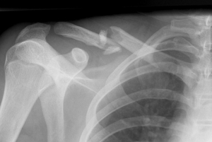
| Read: |
|---|
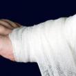
New
- Lady's hand kissing allowance
- Summary of the lesson "first aid for bruises and fractures"
- Food Sources of Ascorbic Acid
- The most dangerous poison for humans
- We study a tree, playing: riddles about a birch
- Plaster cast care
- How to moisturize dry skin in the winter
- Fish oil Biafishenol: instructions for use
- How to clean the apartment yourself from damage
- Amanita mushroom - healing properties, how to take tinctures and prepare ointment


