Sections of the site
Editor's Choice:
- Heart transplant: the possibilities of modern surgery
- Types and reasons for taking a Coombs test
- Endocrine system Neurohypophysis histology
- Offset m echo what. Echoencephalography. For what diseases do echoencephalography
- Nephrotoxic effect of X-ray contrast agents
- Clinical anatomy of the anterior mediastinum Mediastinum: boundaries, division into departments
- Tinnitus causes, diagnosis and treatment
- Baralgin injection: instructions for use Baralgin injections instructions for use reviews
- What a stunned person feels
- Sympathetic and parasympathetic nervous system
Advertising
| What is the human parasympathetic nervous system? The sympathetic and parasympathetic parts of the nervous system. Sympathetic nervous system reactions |
|
According to the morphofunctional classification, the nervous system is divided into: somaticand vegetative.
Somatic nervous systemprovides the perception of irritations and the implementation of motor reactions of the body as a whole with the participation of skeletal muscles. Autonomic nervous system (ANS) innervates all internal organs (cardiovascular system, digestion, respiration, genitals, secretions, etc.), smooth muscles of hollow organs, regulates metabolic processes, growth and reproduction Autonomic (autonomic) nervous system regulates the functions of the body regardless of the will of the person. The parasympathetic nervous system is the peripheral part of the autonomic nervous system, responsible for maintaining the constancy of the internal environment of the body. The parasympathetic nervous system consists of: From the cranial section, in which the preganglionic fibers leave the mid and rhomboid brain as part of several cranial nerves; and From the sacral section, in which the preganglionic fibers leave the spinal cord as part of its ventral roots. The parasympathetic nervous system inhibits work of the heart, dilates some blood vessels. The sympathetic nervous system is a peripheral part of the autonomic nervous system that mobilizes the body's resources for urgent work. The sympathetic nervous system stimulates the heart, constricts blood vessels and enhances skeletal muscle performance. The sympathetic nervous system is represented by: Gray matter of the lateral horns of the spinal cord; Two symmetrical sympathetic trunks with their ganglia; Inter-nodal and connecting branches; and Branches and ganglia involved in the formation of nerve plexuses. All vegetative NS consists of: parasympathetic and sympathetic divisions. Both of these departments innervate the same organs, often exerting the opposite effect on them. The mediator acetylcholine is released by the endings of the parasympathetic part of the autonomic NS. Parasympathetic division of the autonomic NSregulates the work of internal organs at rest. Its activation contributes to a decrease in the frequency and strength of heart contractions, a decrease in blood pressure, an increase in both motor and secretory activity of the digestive tract. The endings of the sympathetic fibers release norepinephrine and adrenaline as mediators. Sympathetic division of the autonomic NS increases its activity if necessarymobilization of body resources. The frequency and strength of heart contractions increase, the lumen of blood vessels narrows, blood pressure rises, the motor and secretory activity of the digestive system is inhibited.
The nature of the interaction between the sympathetic and parasympathetic divisions of the nervous system1. Each of the divisions of the autonomic nervous system can exert an exciting or inhibitory effect on one or another organ. For example, under the influence of sympathetic nerves, the heartbeat increases, but the intensity of intestinal motility decreases. Under the influence of the parasympathetic department, the heart rate decreases, but the activity of the digestive glands increases. 2. If any organ is innervated by both parts of the autonomic nervous system, then their action is usually exactly the opposite. For example, the sympathetic section enhances the contractions of the heart, and the parasympathetic one weakens; the parasympathetic increases the secretion of the pancreas, and the sympathetic decreases. But there are exceptions. So, the secretory nerves for the salivary glands are parasympathetic, while the sympathetic nerves do not inhibit salivation, but cause the release of a small amount of thick viscous saliva. 3. For some organs, either sympathetic or parasympathetic nerves are predominantly suitable. For example, sympathetic nerves go to the kidneys, spleen, sweat glands, and mainly parasympathetic nerves to the bladder. 4. The activity of some organs is controlled by only one part of the nervous system - the sympathetic. For example: when the sympathetic part is activated, sweating increases, and when the parasympathetic part is activated, it does not change, the sympathetic fibers increase the contraction of the smooth muscles that raise the hair, and the parasympathetic ones do not change. Under the influence of the sympathetic part of the nervous system, the activity of some processes and functions can change: blood coagulation accelerates, metabolism occurs more intensively, and mental activity increases. Sympathetic nervous system reactionsThe sympathetic nervous system, depending on the nature and strength of the stimuli, responds either with the simultaneous activation of all its departments, or with reflex responses of individual parts. Simultaneous activation of the entire sympathetic nervous system is most often observed when the hypothalamus is activated (fright, fear, unbearable pain). The result of this massive body-wide response is the stress response. In other cases, reflexively and with the involvement of the spinal cord, certain parts of the sympathetic nervous system are activated. The simultaneous activation of most parts of the sympathetic system helps the body to perform unusually large muscle work. This is facilitated by an increase in blood pressure, blood flow in working muscles (with a simultaneous decrease in blood flow in the gastrointestinal tract and kidneys), an increase in metabolic rate, glucose concentration in blood plasma, breakdown of glycogen in the liver and muscles, muscle strength, mental performance, blood clotting rate ... The sympathetic nervous system is highly excited in many emotional states. In a state of rage, the hypothalamus is stimulated. Signals are transmitted through the reticular formation of the brain stem to the spinal cord and cause a massive sympathetic discharge; all of the above reactions are triggered immediately. This reaction is called the sympathetic anxiety response, or the fight or flight response. an instant decision is required - to stay and engage or flee. Examples of reflexes of the sympathetic nervous system are: - expansion of blood vessels with local muscle contraction; The modified sympathetic ganglion is the adrenal medulla. It produces the hormones adrenaline and norepinephrine, the points of application of which are the same target organs as for the sympathetic part of the nervous system. The action of hormones of the adrenal medulla is more pronounced than that of the sympathetic division. Parasympathetic system responsesThe parasympathetic system carries out local and more specific control of the functions of the effector (executive) organs. For example, parasympathetic cardiovascular reflexes usually act only on the heart, increasing or decreasing the heart rate. Other parasympathetic reflexes act in the same way, causing, for example, salivation or secretion of gastric juice. The emptying reflex of the rectum does not cause any changes over a significant length of the colon. Differences in the influence of the sympathetic and parasympathetic divisions of the autonomic nervous system are due to the peculiarities of their organization. Sympathetic postganglionic neurons have an extensive area of \u200b\u200binnervation, and therefore their excitation usually leads to generalized (broad-acting) reactions. The general effect of the influence of the sympathetic section is to inhibit the activity of most internal organs and stimulate the heart and skeletal muscles, i.e. in preparing the body for behavior such as "fight" or "flight". Parasympathetic postganglionic neurons are located in the organs themselves, innervate limited areas, and therefore have a local regulatory effect. In general, the function of the parasympathetic department is to regulate processes that ensure the restoration of body functions after vigorous activity. The parasympathetic nervous system narrows the bronchi, slows down and weakening heart contractions; narrowing of the blood vessels of the heart; replenishment of energy resources (synthesis of glycogen in the liver and enhancement of digestion processes); strengthening the processes of urination in the kidneys and ensuring the act of urination (contraction of the muscles of the bladder and relaxation of its sphincter), etc. The parasympathetic nervous system predominantly exerts triggering effects: constriction of the pupil, bronchi, activation of the activity of the digestive glands, etc. The activity of the parasympathetic division of the autonomic nervous system is aimed at the current regulation of the functional state, at maintaining the constancy of the internal environment - homeostasis. The parasympathetic department ensures the restoration of various physiological parameters, sharply altered after intense muscular work, and the replenishment of consumed energy resources. The mediator of the parasympathetic system, acetylcholine, reduces the sensitivity of adrenergic receptors to the action of adrenaline and norepinephrine, and has a certain anti-stress effect. Fig. 6. Vegetative reflexes Effect of body position on heart rate (bpm). (Po.Mogendovich M.R., 1972) 3.6.4. Vegetative reflexesThrough autonomic sympathetic and parasympathetic pathways, the central nervous system carries out some autonomic reflexes, starting with various receptors of the external and internal environment: viscero-visceral (from internal organs to internal organs - for example, the respiratory-cardiac reflex); dermo-visceral (from the skin - a change in the activity of internal organs when irritating active points of the skin, for example, acupuncture, acupressure); from the receptors of the eyeball - Ashner's eye-cardiac reflex (slowing down of heartbeats when pressing on the eyeballs - a parasympathetic effect); motor-visceral, for example, orthostatic test (increased heart rate when moving from a lying position to a standing position - a sympathetic effect), etc. (Fig. 6). They are used to assess the functional state of the body and especially the state of the autonomic nervous system (assessing the influence of the sympathetic or parasympathetic department). 11. CONCEPT ABOUT NERVO-MUSCULAR (MOTOR) APPARATUS. MOTOR UNITS (DE) AND THEIR CLASSIFICATION. FUNCTIONAL FEATURES OF DIFFERENT TYPES DE AND THEIR CLASSIFICATION. FUNCTIONAL FEATURES OF DIFFERENT TYPES OF DE. (ACTIVATION THRESHOLD, SPEED AND STRENGTH OF REDUCTION, FATIGUE AND DR) The value of type DE for various types of muscle activity. 12. Muscle composition. Functionality of different types of muscle fibers (slow and fast). Their role in the manifestation of muscle strength, speed and endurance. One of the most important characteristics of skeletal muscles that affect the strength of contraction is the composition (composition) of muscle fibers. There are 3 types of muscle fibers - slow fatigue (type I), fast fatigued or intermediate (type 11-a) and fast fatigued (type 11-b). Slow fibers (type 1), they are also denoted SO - Slow Oxydative (English - slow oxidative) - these are hardy (tireless) and easily excitable fibers, with a rich blood supply, a large number of mitochondria, myoglobin reserves and using oxidative energy production processes (aerobic). There are, on average, 50% of them in humans. They easily engage in work at the slightest muscle tension, are very hardy, but do not have sufficient strength. They are most often used when maintaining non-load static work, for example, while maintaining a pose. Fast fatigued fibers (11-b type) or FG - Fast Glicolitic (fast glycolytic) use anaerobic processes of energy production (glycolysis). They are less excitable, turn on under heavy loads, and provide fast and powerful muscle contractions. But these fibers get tired quickly. There are about 30% of them. Fibers of the intermediate type (P-a) are fast, indefatigable, oxidizing, about 20% of them. On average, different muscles are characterized by a different ratio of slow fatigue and fast fatigue fibers. Thus, in the triceps muscle of the shoulder, fast fibers (67%) predominate over slow ones (33%), which provides the speed-strength capabilities of this muscle (Fig. 14), and the slower and more enduring soleus muscle is characterized by the presence of 84% of slow and only 16 % fast fibers (Saltan B., 1979). However, the composition of muscle fibers in the same muscle has huge individual differences, depending on the innate typological characteristics of a person. By the time a person is born, his muscles contain only slow fibers, but under the influence of nervous regulation, a genetically specified individual ratio of muscle fibers of different types is established in the course of ontogenesis. With the transition from adulthood to the elderly, the number of fast fibers in a person decreases markedly and, accordingly, muscle strength decreases. For example, the largest number of fast fibers in the outer head of the 4th head of the thigh muscle of a man (about 59-63%) is observed at the age of 20-40 years, and at the age of 60-65 years, their number is almost 1/3 less (45%) ...
Fig. 14. The composition of muscle fibers in different muscles Slow ones - in black; fast - gray The number of these or those muscle fibers does not change during training. It is only possible to increase the thickness (hypertrophy) of individual fibers, as well as some change in the properties of intermediate fibers. When the training process is focused on the development of strength, the volume of fast fibers increases, which ensures an increase in the strength of the trained muscles. The nature of nerve impulses changes the strength of muscle contraction in three ways: The mechanical conditions of muscle work are essential - the point of application of its force and the point of application of resistance (lifted load). For example, when flexing at the elbow, the weight of the lifted load can be of the order of 40 kg or more, while the strength of the flexor muscles reaches 250 kg, and the tendon thrust is 500 kg. There is a certain ratio between the force and the speed of muscle contraction, which has the form of hyperbole (the ratio of force - speed, according to A. Hill). The higher the force developed by the muscle, the lower the rate of its contraction, and vice versa, as the rate of contraction increases, the magnitude of the effort decreases. The muscle that works without load develops the highest speed. The speed of muscle contraction depends on the speed of movement of the transverse bridges, that is, on the frequency of stroke movements per unit of time. In fast MUs, this frequency is higher than in slow MUs, and, accordingly, more ATP energy is consumed. During the contraction of muscle fibers in 1 s, about 5 to 50 cycles of attachment-detachment of transverse bridges occur. At the same time, no fluctuations in force in the whole muscle are felt, since MUs work asynchronously. Only with fatigue does the MU work synchronously, and tremors (tremors of fatigue) appear in the muscles. 13. SINGLE AND THETHANIC MUSCLE FIBER REDUCTION. ELECTROMYOGRAM. With a single suprathreshold stimulation of the motor nerve or the muscle itself, the excitation of the muscle fiber is accompanied by single cut. This form of mechanical reaction consists of 3 phases: a latent or latent period, a contraction phase and a relaxation phase. The shortest phase is the latent period, when electromechanical transmission occurs in the muscle. The relaxation phase is usually 1.5-2 times longer than the contraction phase, and when tired it is delayed for a considerable time. If the intervals between nerve impulses are shorter than the duration of a single contraction, then the phenomenon of superposition occurs - the imposition of the mechanical effects of the muscle fiber on each other and a complex form of contraction is observed - tetanus. There are 2 forms of tetanus - dentate tetanus, which occurs with more rare stimuli, when each next nerve impulse enters the relaxation phase of individual single contractions, and solid or smooth tetanus, which occurs with more frequent irritation, when each next impulse falls into the contraction phase ( fig. 11). Thus, (within some boundaries) between the frequency of the excitation pulses and the amplitude of the MU fibers contraction, there is a certain ratio: at a low frequency (for example, 5-8 pulses in 1s)
Fig. P. Single contraction, serrated and solid tetanus soleus muscle human (after: Zimkin N.V. et al., 1984). The upper curve is muscle contraction, the lower curve is the mark muscle irritation on the right is the frequency annoyancesi am single contractions occur, with increasing frequency (15-20 impulses in 1 s) - dentate tetanus, with further increase in frequency (25-60 impulses in 1 s) - smooth tetanus. A single contraction is weaker and less tiring than tetanic. But tetanus provides several times more powerful, albeit short-term, muscle fiber contraction. The contraction of an entire muscle depends on the form of contraction of individual MUs and their coordination in time. When providing long-term, but not very intense work, individual MUs contract alternately (Fig. 12), maintaining the overall muscle tension at a given level (for example, when running long and super-long distances). In this case, individual MUs can develop both single and tetanic contractions, which depends on the frequency of nerve impulses. Fatigue in this case develops slowly, since, working in turn, the DEs manage to recover in the intervals between activation. However, for a powerful short-term effort (for example, lifting a barbell), synchronization of the activity of individual MUs is required, i.e., the simultaneous excitation of almost all MUs. This, in turn, requires simultaneous activation
Fig. 12. Various modes of operation of motor units(DE) the corresponding nerve centers and is achieved through prolonged training. In this case, a powerful and very tedious tetanic contraction is carried out. The amplitude of contraction of a single fiber does not depend on the strength of the suprathreshold stimulation (“All or nothing” law). In contrast, with an increase in the strength of the suprathreshold stimulation, the contraction of the whole muscle gradually increases to its maximum amplitude. Muscle work with a small load is accompanied by a rare frequency of nerve impulses and the involvement of a small number of MUs. Under these conditions, by placing lead electrodes on the skin above the muscle and using amplifying equipment, it is possible to register single action potentials of individual DEs on an oscilloscope screen or using ink recording on paper. In the case of significant voltages, the action potentials of many DEs are algebraically summed up and a complex integrated the trace of recording the electrical activity of the whole muscle is an electromyogram (EMG). The EMG shape reflects the nature of muscle work: under static efforts, it has a continuous form, and with dynamic work - the form of separate bursts of impulses, timed mainly to the initial moment of muscle contraction and separated by periods of "electric silence". The rhythmicity of the appearance of such packs is especially good in athletes during cyclic work (Fig. 13). In young children and individuals not adapted to such work, clear periods of rest are not observed, which reflects insufficient relaxation of the muscle fibers of the working muscle. The greater the external load and the force of muscle contraction, the higher the amplitude of its EMG. This is due to an increase in the frequency of nerve impulses, the involvement of a larger number of units in the muscle and synchronization
Fig. 13. Electromyogram of antagonist muscles during cyclic work their activities. Modern multichannel equipment allows simultaneous registration of EMG of many muscles on different channels. When an athlete performs complex movements, one can see on the EMG curves not only the nature of the activity of individual muscles, but also evaluate the moments and the order of their inclusion or shutdown in different phases of motor acts. EMG records obtained in natural conditions of motor activity can be transmitted to the recording equipment by telephone or radiotelemetry. Analysis of the frequency, amplitude and shape of EMG (for example, using special computer programs) allows one to obtain important information about the peculiarities of the technique of the performed sports exercise and the degree of its mastering by the examined athlete. With the development of fatigue with the same amount of muscle effort, the EMG amplitude increases. This is due to the fact that a decrease in the contractile ability of tired MUs is compensated by the nerve centers by the involvement of additional MUs in the work, i.e., by increasing the number of active muscle fibers. In addition, the synchronization of the MU activity is enhanced, which also increases the amplitude of the total EMG. 14. The mechanism of contraction and relaxation of muscle fibers. Slip theory. The role of the sarcoplasmic reticulum and calcium ions in contraction. With an arbitrary internal command, a person's muscle contraction begins in about 0.05 s (50 ms). During this time, the motor command is transmitted from the cerebral cortex to the motor neurons of the spinal cord and along the motor fibers to the muscle. Approaching the muscle, the excitation process must, with the help of a transmitter, overcome the neuromuscular synapse, which takes about 0.5 ms. The mediator here is acetylcholine, which is contained in synaptic vesicles in the presynaptic part of the synapse. The nerve impulse causes the movement of synaptic vesicles to the presynaptic membrane, their emptying and the release of the mediator into the synaptic cleft. The action of acetylcholine on the postsynaptic membrane is extremely short-lived, after which it is destroyed by acetylcholinesterase to acetic acid and choline. As it is consumed, the reserves of acetylcholine are constantly replenished by synthesizing it in the presynaptic membrane. However, with very frequent and prolonged impulses of the motor neuron, the consumption of acetylcholine exceeds its replenishment, and the sensitivity of the postsynaptic membrane to its action also decreases, as a result of which the conduction of excitation through the neuromuscular synapse is disrupted. These processes underlie the peripheral mechanisms of fatigue during prolonged and heavy muscular work. The released synaptic cleft mediator attaches to the receptors of the postsynaptic membrane and causes depolarization in it. A small subthreshold irritation causes only local excitation of a small amplitude - the potential of the end plate (EPP). With a sufficient frequency of nerve impulses, the EPP reaches a threshold value and a muscular action potential develops on the muscle membrane. It (at a speed of 5) spreads along the surface of the muscle fiber and enter the transverse tubules inside the fiber. By increasing the permeability of cell membranes, the action potential causes the release of Ca ions from the cisterns and tubules of the sarcoplasmic reticulum, which penetrate into myofibrils, to the binding centers of these ions on actin molecules. Under the influence of Sadlinny tropomyosin molecules turn along the axis and hide in the grooves between the spherical actin molecules, opening the sites of attachment of the myosin heads to actin. Thus, so-called cross bridges are formed between actin and myosin. In this case, the myosin heads perform rowing movements, ensuring the sliding of the actin filaments along the myosin filaments from both ends of the sarcomere to its center, ie, the mechanical reaction of the muscle fiber (Fig. 10). The energy of the rowing motion of one bridge produces a movement of 1% of the length of the actin filament. For further sliding of contractile proteins relative to each other, the bridges between actin and myosin must disintegrate and re-form at the next Ca-binding site. This process occurs as a result of the activation of myosin molecules at this moment. Myosin acquires the properties of the enzyme ATPase, which causes the breakdown of ATP. The energy released during the decay of ATP leads to the destruction
Fig. 10. Diagram of the electromechanical connection in the muscle fiber On A: a state of rest, on B - arousal and contraction yes - action potential, mm - muscle fiber membrane, n _ transverse tubes, t - longitudinal tubes and cisterns with ions Ca, a - thin filaments of actin, m - thick filaments of myosin with bulges (heads) at the ends. Zeta membranes are limited sarcomeres of myofibrils. Thick arrows - potential spread actions during fiber excitation and movement of Cais ions in tanks and longitudinal tubules into myofibrils, where they promote the formation bridges between the filaments actin and myosin and the sliding of these filaments (fiber contraction) due to the rowing movements of the myosin heads. existing bridges and the formation in the presence of Sanovye bridges in the next section of the actin filament. As a result of the repetition of such processes of multiple formation and disintegration of bridges, the length of individual sarcomeres and the entire muscle fiber as a whole is reduced. The maximum concentration of calcium in the myofibril is reached within 3 ms after the appearance of the action potential in the transverse tubules, and the maximum tension of the muscle fiber - after 20 ms. The whole process from the appearance of a muscle action potential to the contraction of a muscle fiber is called electromechanical coupling (or electromechanical coupling). As a result of muscle fiber contraction, actin and myosin are more evenly distributed inside the sarcomere, and the transverse striation of the muscle, visible under the microscope, disappears. Relaxation of the muscle fiber is associated with the work of a special mechanism - the "calcium pump", which ensures the pumping of Cais ions of myofibrils back into the tubules of the sarcoplasmic reticulum. This also consumes the energy of ATP. 15. The mechanism of regulation of the strength of muscle contraction (the number of active MUs, the frequency of impulses of motoneurons, synchronization of the contraction of muscle fibers of different MUs in time). The nature of nerve impulses changes the strength of muscle contraction in three ways: 1) an increase in the number of active MUs is a mechanism for the involvement or recruitment of MUs (first, slow and more excitable MUs are involved, then - high-threshold fast MUs); 2) an increase in the frequency of nerve impulses, as a result of which there is a transition from weak single contractions to strong tetanic contractions of muscle fibers; 3) an increase in the synchronization of the MU, while there is an increase in the contraction force of the whole muscle due to the simultaneous traction of all active muscle fibers. The parasympathetic nervous system "balances" the sympathetic. It ensures the adaptation of the eyes to vision at close range, a decrease in the heart rate, the activation of the secretion of saliva and other digestive juices, as well as an increase in intestinal motility. The most striking example of the coordinated activity of the parasympathetic and sympathetic systems is their interaction during intercourse. The central part of the parasympathetic nervous system consists of the head (cranial) section and the spinal (sacral) section. Preganglionic fibers extend from the brain stem as part of the four cranial nerves (oculomotor, facial, glossopharyngeal, and vagus) and from the sacral segments of the spinal cord. The structure of the parasympathetic nervous system (ganglionic neurons and postganglionic fibers are highlighted in red).and) Cranial parasympathetic system... Preganglionic fibers are distributed in the four cranial nerves: 1. As part of the oculomotor nerve, which forms a synapse with the ciliary ganglion. Postganglionic fibers are responsible for the innervation of the muscles involved in the accommodation reflex - the sphincter of the pupil and ciliary muscle. 2. As part of the facial nerve, which forms a synapse with the pterygopalatine ganglion (responsible for the innervation of the lacrimal and nasal glands) and the submandibular ganglion (responsible for the innervation of the submandibular and sublingual salivary glands). 3. As part of the glossopharyngeal nerve, which forms a synapse with the auricular ganglion (responsible for innervation). 4. As part of the vagus nerve, which forms synapses with extramural (located near the innervated organ) and with intramural (located in the wall of the innervated organ) ganglia of the heart, lungs, lower esophagus, stomach, pancreas, gallbladder, small intestine, as well as the ascending and the transverse colon.  The cranial section of the parasympathetic system. E-V-core of Edinger-Westphal; ZYABN - the posterior nucleus of the vagus nerve. The explanation of the rest of the abbreviations is presented below the figure above (here we will duplicate them). The cranial section of the parasympathetic system. E-V-core of Edinger-Westphal; ZYABN - the posterior nucleus of the vagus nerve. The explanation of the rest of the abbreviations is presented below the figure above (here we will duplicate them). RH-ciliary ganglion; SG-cardiac ganglia; IG-intramural ganglia; MG-myenteric ganglia (ganglia associated with the muscular layer of the intestine); UG ear ganglion; TG-pelvic ganglia; KG-pterygopalatine ganglion; PG-submandibular ganglion. b) Sacral section of the parasympathetic system... Behind the first lumbar vertebra, the sacral segments of the spinal cord form its terminal part - the cerebral cone of the spinal cord. The gray matter of the lateral horns of the sacral segments S2, S3 and S4 of the spinal cord gives rise to preganglionic fibers, which, propagating caudally in the anterior roots of the spinal cord, pass into the cauda equina. After exiting the pelvic sacral foramen, some of the fibers branch off and form the pelvic visceral nerves. Fibers of the left and right visceral pelvic nerves form synapses either with ganglion cells located in the walls of the colon (distal) and rectum, or with the pelvic parasympathetic ganglia adjacent to the pelvic sympathetic ganglia described above. Postganglionic parasympathetic fibers are responsible for the innervation of the bladder detrusor, as well as the middle membrane of the internal pudendal artery and its branches, going to the cavernous tissue of the clitoris or penis. Autonomic Nervous System (ANS) Anatomy Instructional Video
The sympathetic department is a part of the autonomic nervous tissue, which, together with the parasympathetic, ensures the functioning of internal organs, chemical reactions that are responsible for the vital activity of cells. But you should know that there is a metasympathetic nervous system, a part of the vegetative structure located on the walls of organs and capable of contracting, contacting directly with the sympathetic and parasympathetic, making adjustments to their activity. The internal environment of a person is directly influenced by the sympathetic and parasympathetic nervous systems. The sympathetic division is localized in the central nervous system. The spinal nerve tissue operates under the control of nerve cells in the brain. All elements of the sympathetic trunk, located two from the spine of the side, are directly connected with the corresponding organs through the nerve plexuses, each with its own plexus. At the bottom of the spine, both trunks in humans are combined together. The sympathetic trunk is usually divided into sections: lumbar, sacral, cervical, thoracic.
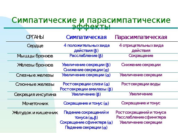
These plexuses are divided into smaller ones, and from them the impulses move to the internal organs. The transition of excitation from the sympathetic nerve to the corresponding organ occurs under the influence of chemical elements - sympatines, secreted by nerve cells. They supply the same tissues with nerves, ensuring their interconnection with the central system, often exerting a direct opposite effect on these organs. The influence that the sympathetic and parasympathetic nervous systems have can be seen from the table below:
If one begins to prevail over the other, symptoms of increased excitability of sympathicotonia appear (the sympathetic part predominates), vagotonia (parasympathetic predominates). Sympathicotonia manifests itself in the following symptoms: fever, tachycardia, numbness and tingling in the extremities, increased appetite without the appearance of being deprived of weight, indifference to life, restless dreams, fear of death for no reason, irritability, absent-mindedness, salivation decreases, as well as sweating, migraine appears. In a person, when the increased work of the parasympathetic department of the vegetative structure is activated, increased sweating appears, the skin is cold and wet to the touch, a decrease in the heart rate occurs, there is less than the prescribed 60 beats in 1 minute, fainting, salivation and respiratory activity increase. People become indecisive, slow, prone to depression, unbearable. The parasympathetic nervous system reduces the activity of the heart, tends to dilate blood vessels. FunctionsThe sympathetic nervous system is a unique design of an element of the autonomic system, which, in the event of a sudden need, is able to increase the body's ability to perform work functions by collecting possible resources. As a result, the structure carries out the work of organs such as the heart, reduces blood vessels, increases the ability of muscles, frequency, heart rate strength, efficiency, inhibits the secretory, absorption capacity of the gastrointestinal tract.
SNS supports functions such as the normal functioning of the internal environment in an active position, being activated during physical effort, stressful situations, diseases, blood loss and regulates metabolism, for example, an increase in sugar, blood clotting, and others. It is most fully activated during psychological shocks, by producing adrenaline (the enhancing action of nerve cells) in the adrenal glands, which enables a person to react faster and more efficiently to sudden factors from the outside world. Also, adrenaline can be produced with increasing load, which also helps a person to better cope with it. After coping with the situation, a person feels tired, he needs to rest, this is due to the sympathetic system, which has most fully used up the body's capabilities, in connection with an increase in body functions in a sudden situation. Parasympathetic NS performs the functions of self-regulation, protection of the body, is responsible for emptying a person. Self-regulation of the body has a restorative effect, working in a calm state. The parasympathetic part of the activity of the autonomic nervous system is manifested by a decrease in the strength and frequency of the heart rate, stimulation of the gastrointestinal tract with a decrease in glucose in the blood, etc. Carrying out protective reflexes, it relieves the human body of foreign elements (sneezing, vomiting, and others). The table below shows how the sympathetic and parasympathetic nervous systems act on the same elements of the body. TreatmentIf you notice signs of increased sensitivity, you should consult a doctor, as this can cause an ulcerative, hypertensive nature, neurasthenia. Only a doctor can prescribe the correct and effective therapy! There is no need to experiment with the body, since the consequences if the nerves are in a state of excitability is a rather dangerous manifestation not only for you, but also for people close to you. When prescribing treatment, it is recommended, if possible, to eliminate the factors that excite the sympathetic nervous system, be it physical or emotional stress. Without this, no treatment, most likely, will help; after drinking a course of medications, you will get sick again. You need a cozy home environment, sympathy and help from loved ones, fresh air, good emotions. First of all, you need to make sure that nothing raises your nerves. The drugs used in the treatment are based on the group of potent drugs, so they should be used with caution only as directed or after consulting a doctor. Prescribed drugs usually include: tranquilizers ("Phenazepam", "Relanium" and others), antipsychotics ("Frenolon", "Sonapax"), hypnotics, antidepressants, nootropic drugs and, if necessary, cardiac ("Korglikon", "Digitoxin" ), vascular, sedative, vegetative preparations, a course of vitamins. It is good when using physiotherapy, including physiotherapy exercises and massage, you can do breathing exercises, swimming. They are good at helping to relax the body. In any case, ignoring the treatment of this disease is strongly discouraged, it is necessary to consult a doctor in a timely manner, to carry out the prescribed course of therapy.
Sympathetic nervous systemCute ANS consists of central and peripheral sections (Fig. 5.1). Central department located in the lateral horns of the spinal cord from the 1st thoracic to the 3rd lumbar segments. Peripheral - consists of nerve fibers and nodes of the paravertebral (bilacular) and prevertebral (anterior). The paravertebral nodes are located segment by segment in two chains on the sides of the spine, forming the right and left sympathetic trunks. Prevertebral nodes are nodes of the peripheral plexuses of the chest and abdominal cavities (abdominal, mesenteric, superior and inferior). Sympathetic nerve fibers leave the spinal cord as part of the anterior roots of the spinal nerves, and then through the preganglionic (prenodal) fibers - the white connecting branch - are sent to the corresponding node (ganglion) of the sympathetic trunk. In it, some fibers pass to the postganglionic (pislyavuzlovy) neuron, which is sent to the organs (blood vessels, sweat glands). The second pass through the sympathetic trunk node without interruption (transit) and enter the prevertebral nodes, switch to them, and then, like postganglionic efferent fibers, stretch to the corresponding organs (lungs and others). There is an opinion that, in addition to efferent fibers, the sympathetic nervous system has its own sensory (afferent) fibers (in the myocardium). Depending on localization FIG. 5.1. cell bodies, course and length of branches, they can be divided into two groups. The first group of peripheral afferent neurons includes cells, the bodies of which are localized in the prevertebral sympathetic nodes. One of the long branches follows to the periphery, the other to the side of the spinal cord, which is part of the dorsal roots. The second group is characterized by the fact that a long branch of these sensitive cells is associated with the working organ. Short branches are distributed in the node itself, synaptically contact with plug-in neurons, and through them - with effector neurons and create a local reflex arc here. Parasympathetic nervous systemThe parasympathetic ANS is also central and peripheral. Central the department consists of parasympathetic nuclei, laid in the middle and medulla oblongata and sacral segments (2-4) of the spinal cord. Peripheral department - nodes and fibers that make up the oculomotor (III pair), facial (VII pair), lingopharyngeal (IX pair), vagus (X pair) nuclei and pelvic nerves. In the midbrain, at the bottom of the aqueduct, there is a parasympathetic additional oculomotor nucleus (the nucleus of Yakubovich - Edinger - Westphal), the processes of the cells of which are directed as part of the oculomotor nerve, switch to the ciliary node (contained in the orbit) and end in the muscle that narrows the pupils, and in the ciliary muscles. The rhomboid fossa next to the nucleus of the facial nerve contains the salivary cranial (superior) nucleus. The processes of its cells are part of the intermediate nerve, then the facial one. Together with the branches of the facial and trigeminal nerves, the parasympathetic fibers reach the lacrimal gland, the glands of the mucous membrane of the nasal and oral cavities (switch in the pterygopalatine node) and the submandibular and sublingual glands (switch in the adjacent submandibular node). The salivary tail (lower nucleus) gives rise to the parasympathetic (secretory) fibers of the parotid gland, which leave the brain as part of the IX pair (lingopharyngeal nerve) and switch in the ear node. The bulk of the parasympathetic fibers that emerge from the medulla oblongata, which are part of the vagus nerve. They start from its parasympathetic dorsal (dorsal) nucleus at the bottom of the rhomboid fossa. The prenodal fibers stretch to the organs of the neck, chest and abdominal cavities, ending in the intramural ganglia (inside organs), nodes of the thyroid and thymus glands, in the bronchi, lungs, heart, esophagus, stomach, intestines, in the pancreas, liver, kidneys. From the intramural nodes, postnodal fibers extend, which innervate these organs. From the sacral segments of the spinal cord, parasympathetic prenodal fibers are directed as part of the ventral roots of the sacral nerves, and when separated from them, they form the internal pelvic nerves. their branches enter the hypogastric plexus and end on the cells of the intramural nodes. The post-nodular fibers innervate the smooth muscles and glands of the lower parts of the digestive system, urinary, external and internal genital organs. The main collector of the sensitive pathways of the parasympathetic nervous system is wandering nerve. Afferent fibers of his cervical spine are 80-90 %. About 20 % of them are myelinated, the rest are thin myelin-free. These fibers transmit information from the digestive tract, chest and abdominal organs. The receptors formed by these fibers react to mechanical, thermal, painful influences, perceive changes in pH and electrolyte composition. The physiological role of the sensitive branch of the vagus nerve is extremely important - depressor nerve. It is a powerful conductor that signals the level of blood pressure in the aorta. The cells of the body of the afferent pathways of the vagus nerve are located mainly in the jugular node, and their fibers enter the medulla oblongata at the level of the olives. As part of the sinus nerve, it is a branch of the IX pair, contains about 300 thick afferent fibers, which are associated with a large number of receptors of various modality. In this perceiving complex, a special role belongs to the carotid glomeruli, which is located between the internal and external carotid arteries at the branching point of the common carotid artery (carotid sinus, sinus caroticus). Thus, the composition of the autonomic nervous system includes: ■ nerve fibers; ■ peripheral nerve ganglia, consisting of nerve cells; ■ below nerve centers - located in the gray matter of the spinal cord and the brain stem, from the cells of which efferent nerve fibers begin; ■ higher nerve centers - located in the diencephalon and forebrain. |


 Together they are responsible for cardiovascular organisms, digestive organs, respiratory structures, secretions, the work of the smooth muscle of the hollow organs, control the metabolic processes, growth, reproduction.

New
- Confused consciousness: symptoms, causes and treatment
- Laparoscopy (removal) of appendicitis
- The liver is not palpable what does it mean Determination of dimensions according to Kurlov
- Pathological types of bite and methods of their correction Tests for determining bite anomalies
- Cancer stages International tumor classification
- Inflammation of the appendages in women - symptoms and treatment regimen
- Thyroid disease in women
- Examination of the spleen. Palpation of the spleen. Spleen palpation technique Spleen along the costal margin
- All about follicles in the ovaries
- The superior vena cava is formed from the fusion of veins


